Author: Sidney Ocanagil-Tunstall
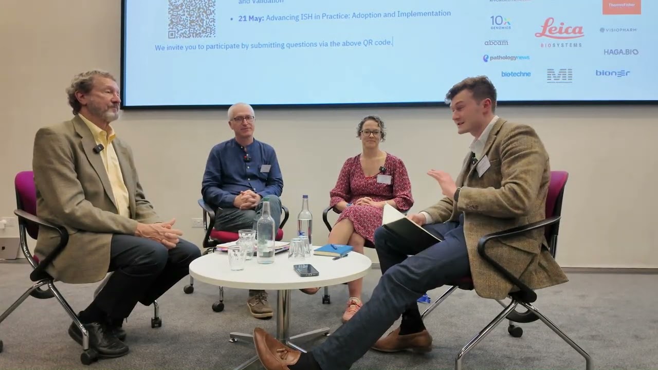
The fourth In Situ Hybridisation and Spatial Omics Symposium, held at the Cancer Research UK Cambridge Institute from 20–21 May 2026 explored how in situ hybridisation (ISH) methods are evolving within the spatial biology landscape. Across the two-day programme, talks covered advances in ISH technologies, disease applications, multiomics, AI and 3D biological systems.
While the meeting showcased expanding technical toolkits for researchers and clinicians, one conversation in particular stood out: spatial biology is now producing images and datasets that are too rich to interpret through conventional methods alone.
This challenge was captured most clearly in Professor John Le Quesne’s talk, “Self-supervised Artificial Intelligence for the Interpretation of Data-Rich Spatial Biology Images.” His group at the CRUK Scotland Institute works with spatial biology and clinical pathology, using technologies including multiplexed RNA and protein detection.
While supervised AI is particularly valuable, it is limited by existing human labels and assumptions and will only work with what it already knows how to define. Self-supervised AI offers something different: a way to learn directly from complex tissue images and uncover patterns that may not yet have a diagnostic significance or biological name.
This matters because spatial biology images now combine morphology, molecular phenotype, cellular neighbourhoods and tissue architecture. Multi-dimensional information, far beyond the single marker haematoxylin and eosin (H&E) stained tissue that has been the backbone of histopathology since the late 19th century. Le Quesne’s group has used self-supervised learning to build histomorphological atlases for lung adenocarcinoma and mesothelioma, identifying previously unrecognised “lethal” morphologies.
Talks on computational phenotyping, tumour microenvironment profiling and AI-driven ISH quantification all pointed towards the same conclusion: as spatial biology becomes more multiplexed and multiomic, our ability to interpret the data becomes the bottleneck.
The last decade has seen the use of AI for automating existing lab tasks and assisting the histopathologist with tumour detection. While there is no doubt that we will continue to see the use of AI in this fashion, this symposium showcased that the AI use case will ultimately move beyond these applications toward detailed interpretation. The next challenge lies in turning the data-rich images, that we now have the capabilities to create, into biological insight, clinically relevant biomarkers, and validated tools that can support real-world translational research.
With the scene now set, can we now say for certain that tools derived from self-supervised AI will ultimately become one of the most important technologies in the researchers and clinician’s toolbox?