Immediate margin assessment
Bringing The Unseen Into Focus For A Cancer-free Tomorrow
Cancer Cells At Your Fingertips
SamanTree Medical was born out of a novel ultra-fast confocal microscopy technology invented at EPFL (Switzerland) in 2010. Since then we have re-invented confocal microscopy to make it compatible with the intraoperative environment.
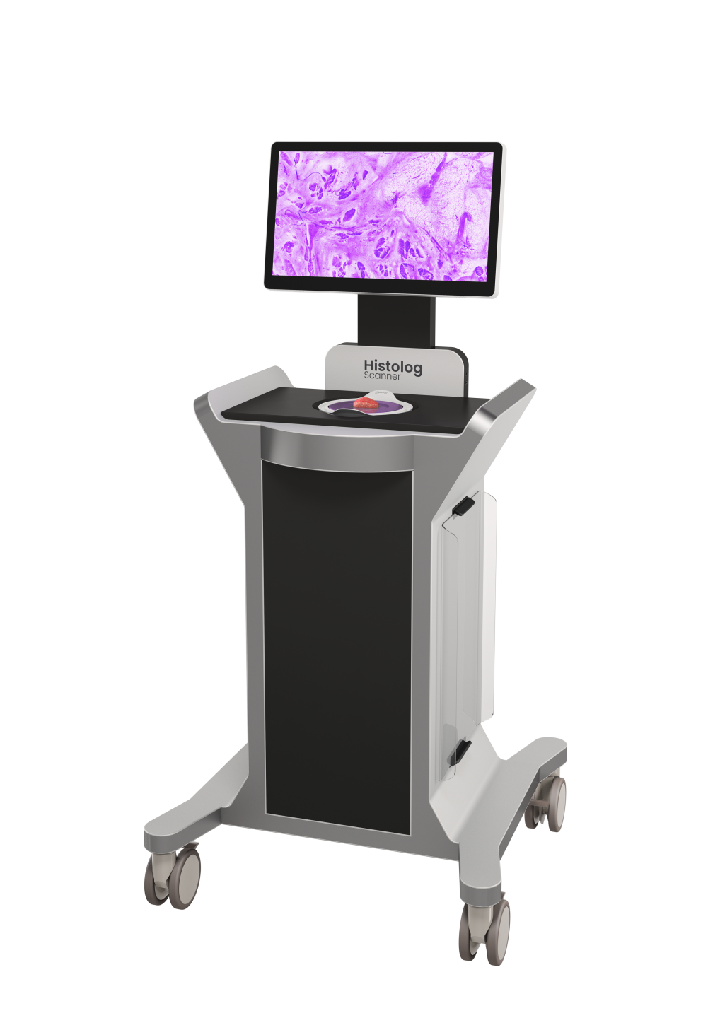
The Histolog® Scanner
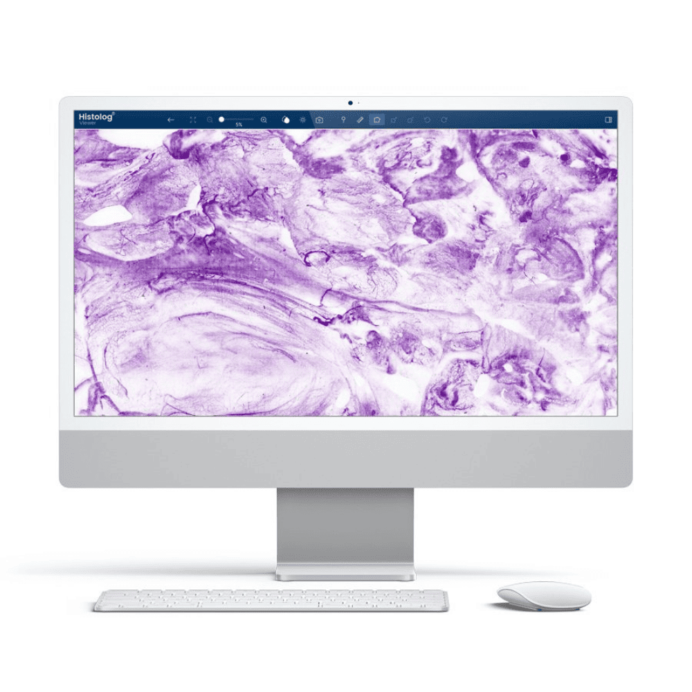
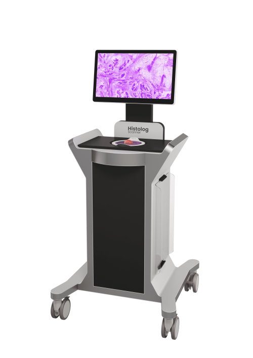
The Histolog Scanner (CE mark since 2018) is a digital microscopy scanner for high-resolution imaging of the surface of fresh tissue. Based on a novel ultra-fast confocal microscopy technology invented in EPFL. Its innovative design makes it highly practical for quick assessment on the point-of-care and the clinician is one touch on the screen away from visualizing cancerous cells immediately on a surgical specimen¹ ⁻ ⁸.
Seamless integration into your clinical routine
10 second specimen preparation time
45 seconds for image acquisition
Remote workflow in a single tap
The Histolog Dip and Dish
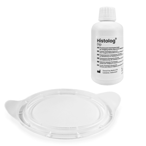
The Histolog Dip (CE-IVD) is a fluorescent histological stain for use as an accessory device in combination with the Histolog Scanner for imaging the surface of excised human tissue specimens to visualize cell morphology. It is designed for rapid imaging of fresh tissue, while preserving specimen integrity for future pathological analysis ² ⁻ ⁵.
The Histolog Dish (CE-IVD) is a single-use protection, preventing direct contact of the tissue specimen with the Histolog Scanner during imaging.

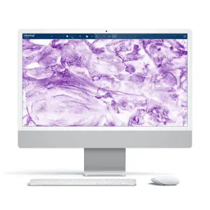
The Histolog Remote
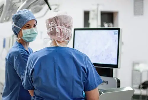
The Histolog Remote features is a collaboration tool that lets pathologists and surgeons work together in real time, even if they’re not in the same room. This allows pathologists to analyze the specimen’s surfaces and provide instant feedback, allowing surgeons to make quicker, more informed decisions in the operating room. It’s a straightforward, efficient way to get expert input, no matter where the team is located.
The Histolog solution has demonstrated effectiveness in applications related to prostate and breast tissues among others
Elfgen C, Papassotiropoulos B, Varga Z, et al. Comparative analysis of confocal microscopy on fresh breast core needle biopsies and conventional histology. Diagn Pathol. 2019;14:58. doi:10.1186/s13000-019-0835-z.
Sandor MF, et al. Imaging of lumpectomy surface with large field-of-view confocal laser scanning microscope for intraoperative margin assessment – POLARHIS study. The Breast. 2022;66:118-125. doi:10.1016/j.breast.2022.10.003.
Sandor MF, et al. Imaging of lumpectomy surface with large field-of-view confocal laser scanning microscope for intraoperative margin assessment – POLARHIS study. The Breast. 2022;66:118-125. doi:10.1016/j.breast.2022.10.003.
Sandor MF, et al. Imaging of lumpectomy surface with large field-of-view confocal laser scanning microscope for intraoperative margin assessment – POLARHIS study. The Breast. 2022;66:118-125. doi:10.1016/j.breast.2022.10.003.
Wernly D, Beniere C, Besse V, et al. SENOSI Confocal Microscopy: A new and innovating way to detect positive margins in non-palpable breast cancer? Life. 2024;14(2):204. doi:10.3390/life14020204.
Mathieu MC, Ragazzi M, Ferchiou M, et al. Breast tissue imaging atlas using ultra-fast confocal microscopy to identify cancer lesions. Virchows Arch. 2025;486:299–311. doi:10.1007/s00428-024-03783-y.
Sandor MF, Schuller Z, Heimann S, et al. SHIELD study: Reoperation rate in breast-conserving surgery using confocal histology scanner for intraoperative margin assessment – Interim results. Thieme. 2024. doi:10.1055/a-2419-6777.
Eissa A, Puliatti S, Rodriguez Peñaranda N, et al. Current applications of ex-vivo fluorescent confocal microscope in urological practice: A systematic review of literature. Chin Clin Oncol. 2024;13(4):52. doi:10.21037/cco-23-150
Windisch O, Diana M, Tilki D, et al. Intraoperative technologies to assess margin status during radical prostatectomy – A narrative review. Prostate Cancer Prostatic Dis. 2025;28:81–88. doi:10.1038/s41391-024-00868-2
Dinneen E, Almeida-Magana R, Al-Hammouri T, et al. Intraoperative margin assessment during radical prostatectomy: Is microscopy frozen in time or ready for digital defrost? Histopathology. 2024;85:716–726. doi:10.1111/his.15290
Almeida-Magana R, Au M, Al-Hammouri T, et al. Accuracy of the LaserSAFE technique for detecting positive surgical margins during robot-assisted radical prostatectomy: Blind assessment and inter-rater agreement analysis. Histopathology. 2025;86:433–440. doi:10.1111/his.15336