
Author: Sidney Ocanagil-Tunstall
This article is part one in our Technology Buyers’ Guide series, ‘Where are you in your digital pathology journey?’ In this article we aim to share the advantages of going digital with lab staff but also make clear that in order for your future digital workflow to prosper it will require a holistic change to your histopathology service.
Pathology’s Physical Constraint
Take a common scenario. Your histopathologist comes across a case that requires a specialist opinion, or he/she would like a second opinion on. The expertise exists, but it sits in another hospital, another region or perhaps even another country. Before that expertise can be brought to bear, the material has to make the journey first.
Someone must locate the glass slides, package them and arrange their transport. Until they arrive, the case waits and so does an anxious patient.
This is one example of a wider constraint built into traditional histopathology. Glass slides are essential diagnostic objects, but they can only be in one place at a time. Meanwhile, pathology services increasingly operate across networks. Specialist expertise is spread between locations and cases often involve contributions from several clinical teams.
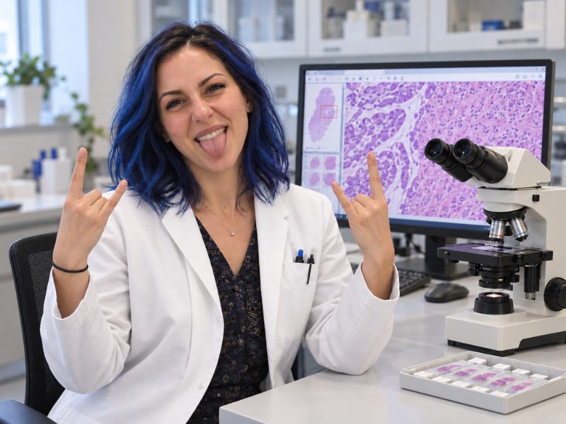
Digital pathology changes that relationship. By scanning glass slides to create high-resolution digital images, laboratories can begin moving information between people and places more easily. Cases can be made available for reporting, consultation, multidisciplinary discussion, teaching and research without every use depending on the physical location of the slide.
The possibilities are considerable, but their value will differ from one laboratory to another. A service struggling to access specialist advice has a different starting point from one trying to share reporting work across several sites. Another may be interested in improving teaching, building research collections or preparing for image analysis and artificial intelligence.
The first question, therefore, is not which scanner to buy. It is: what could digital pathology allow your laboratory to do differently?
This article begins the Pathology News Technology Buyers’ Guide buying journey. It explores the opportunities digital pathology can create before the journey moves on to defining the problem, assessing readiness and evaluating the technologies that might help.
From Moving Slides to Moving Information
In clinical histopathology, digital pathology commonly centres on whole slide imaging. A scanner captures a high-resolution digital representation of the tissue on a glass slide. A pathologist can then navigate that image on screen, moving from an overview of the specimen to areas requiring closer examination.
The glass slide remains. Tissue must still be processed, sectioned, stained and mounted to a standard suitable for its intended use. By digitising, we are adding an additional stage to that pathway: scanning, checking, managing and presenting the image.
Three parts of this process are useful to distinguish:
- Scanning covers scanning the slide and creating the digital image.
- Image management and workflow connect that image to the correct case, store it and make it available alongside the information needed for review.
- Image analysis and AI provide additional computational capabilities for particular tasks.
A laboratory can use digital slides for reporting, consultation or teaching without introducing AI. Equally, installing a scanner does not create a complete digital service.
Imagine following a case through the workflow. Once the physical slide is ready, it is identified and scanned. The resulting image is checked to establish whether the tissue has been captured adequately and is sufficiently in focus. If there is a problem, staff need to know whether the slide should be rescanned, reprocessed or returned to the pathologist for review by glass.
A suitable image then needs to reach the right case. The pathologist must be able to open it with the relevant clinical information, review it with suitable performance and complete the report through the laboratory’s established systems.
The value of digital pathology rests on this complete chain. The decisive moment is not when the scanner captures an image. It is when the right image opens beside the right case, at the right quality, when someone needs to use it.
The Technology Buyers’ Guide connects these steps to categories including Pre-analytics, Slide Scanning, Image Quality Control, LIMS & Tracking Systems, Data Storage, IMS & PACS, and Image Analysis. Exploring the workflow through these categories can help you understand the role each technology plays and how the parts need to work together.
What Becomes Possible When Location Matters Less?
Perhaps the clearest opportunity created by digital pathology is easier access to expertise.
Consider a general pathologist reviewing an unusual skin lesion. An agreed digital consultation pathway could allow a dermatopathologist at another site to access the images and relevant case information. Both colleagues can examine the same material, with annotations helping to draw attention to the area that prompted the referral.
The same capability can support multidisciplinary team meetings. Instead of pathology being represented only through the written report, relevant histological features can be displayed and discussed as part of the wider clinical picture.
Digital access can also give networks more options for distributing work. If one site has a growing queue while another has appropriate reporting capacity, suitable cases may be allocated across the network. Remote reporting arrangements may allow pathologists to access work from another hospital or an approved home workstation.
Consultation, remote diagnosis and the distribution of diagnostic work are among the use cases described in the Leeds Guide to Digital Pathology.
The potential is significant: laboratories can begin organising work around the availability of expertise rather than the location of physical slides.
Yet access alone does not create capacity. A specialist still needs time to examine the case, and a network needs clear arrangements for allocating work, accepting responsibility and returning the report. Digital pathology gives services more freedom to organise the expertise they already have; whether that improves turnaround depends on how the wider service operates.
Pathology News’ laboratory showcases, interviews and Thought Leaders discussions explore how laboratories have approached these organisational questions in practice. When reading them, look beyond the installed technology. Who allocates cases? How do colleagues communicate? Who takes responsibility when an image cannot be reported digitally? These decisions often reveal more about a working service than the equipment list alone.
Giving Existing Material a New Level of Usefulness
The value of a digital image does not end with its first review.
A previous biopsy may help a pathologist interpret a current specimen. In a physical workflow, accessing it can involve an archive search, retrieval and delivery to the reporting room. Where the material has already been scanned and correctly linked to the patient and case, it may be available electronically.
Easier access to previous slides is one of the potential benefits identified by Leeds Teaching Hospitals. Its value will depend on local demand. A laboratory could begin by asking how often previous material is requested, which cases generate the most requests and how much time is spent retrieving it.
An existing glass archive does not become digitally available when a laboratory starts scanning new work. Services need to decide whether selected historical material should be digitised, when that should happen and how the resulting images will be organised and retained.
Once images are available, they may also support peer review and quality improvement. Colleagues can examine the same material, return to a particular region and discuss differences in interpretation. Teams can review recurring preparation or scanning problems using the affected images alongside records from the workflow.
Imagine a laboratory experiencing frequent requests to rescan slides. Bringing those cases together may reveal a repeated issue with tissue placement, staining, coverslipping or the scanning process itself. The image then becomes more than a diagnostic representation; it becomes evidence that can help the team understand and improve its own workflow.
Reliable identification and tracking support this work by helping staff establish which slide belongs to which case and what happened as it moved through the laboratory. Digitisation does not eliminate identification errors, but a well-designed workflow can make activity more visible and easier to investigate.
The Buyers’ Guide’s Pre-analytics and Image Quality Control categories provide a useful place to explore this relationship. Looking at them together reinforces an essential point: the quality of a digital image begins long before the slide reaches the scanner.
More Than a Reporting Tool
Once pathology material can be accessed digitally, its usefulness can extend into education and research.
A teaching case is often valuable to more than one trainee and on more than one occasion. A digital collection can allow learners across different locations to review the same material, mark areas of uncertainty and return to those regions with a supervisor. Rather than sharing a microscope or waiting for a slide set, trainees can work through cases independently and use their time with an expert for a more focused discussion.
The educational value still comes from the selection and explanation of the cases. A folder of images is not a teaching programme. Collections need to be curated around defined learning needs, supported by relevant information and managed with appropriate access arrangements.
Research introduces a different set of possibilities. Digital images can support review and annotation across defined groups of cases, collaboration between authorised teams and the development or evaluation of computational tools.
The National Pathology Imaging Co-operative provides one example of this wider potential. Its infrastructure supports high-volume scanning, dataset curation, secure data access, algorithm evaluation, technical validation and research into how digital systems interact with clinical practice.
A clinical archive, however, is not automatically a research-ready dataset. Researchers need to establish whether the images are suitable, whether the accompanying information is sufficiently complete and whether the proposed use has the appropriate governance and permissions.
For laboratories with academic or industry research partners, considering these future uses early can influence how images and associated information are stored and organised. It can also prevent a familiar problem: building a large collection only to discover that it lacks the consistency or metadata needed to answer the research question.
Pathology News’ Deeper Insights, Thought Leaders interviews and research coverage can help readers examine how clinical infrastructure is being used beyond routine diagnosis—and where the gap remains between technical possibility and useful evidence.
Opening the Door to Image Analysis and AI
Digital slides also create the foundation on which image-analysis and AI tools operate.
Depending on the intended application, software may help measure structures, quantify particular features or highlight regions for review. These capabilities could support tasks that are repetitive, time-consuming or subject to variation between observers.
The most useful starting point is a specific activity. What part of the current assessment is difficult? What would the software contribute? How would its output reach the pathologist, and what decision would it inform?
Without that clarity, AI risks becoming a broad ambition in search of a problem.
Performance also needs to be considered in the setting where the tool will be used. Specimen preparation, staining, image quality, case mix and scanner characteristics may all affect its behaviour. Results reported in a research study do not automatically demonstrate value in routine clinical practice.
Digital pathology can deliver useful capabilities before AI enters the workflow. Easier consultation, flexible access and improved retrieval may provide an immediate reason to digitise, while computational tools are assessed separately against defined needs.
The Buyers’ Guide’s Image Analysis category allows readers to explore the tasks individual solutions are intended to address. Pathology News’ AI reporting, podcasts, webinars and expert discussions provide further context around the evidence, implementation and clinical questions surrounding their use.
The Reality Behind the Promise
The possibilities of digital pathology ultimately meet a simple operational test: does the service work when people depend on it?
Good tissue preparation remains fundamental. Scanning and image-quality checks must support the intended application. Images and case information need to be connected reliably, while storage and network infrastructure must deliver suitable access and viewing performance.
Responsibilities need to be equally clear. Who monitors the scanners? Who reviews image quality? Who manages exceptions? What happens if a system is unavailable? How does a pathologist request the glass slide when digital review is insufficient?
Clinical use also requires appropriate assurance. Pathologists need suitable training and validation, and laboratories must consider the professional, regulatory and accreditation requirements applying to their service. The College of American Pathologists’ whole slide imaging guideline provides one professional reference for validating intended diagnostic applications in the US. Laboratories elsewhere should establish the requirements relevant to their own jurisdiction.
Planning must extend beyond the initial purchase. Software, support, maintenance, storage growth, staff time and continuity arrangements all shape the long-term service.
These are not peripheral technical details. Each one determines whether the theoretical benefit reaches the reporting pathologist and, ultimately, the patient.
The Pathology News NYU Langone laboratory showcase explores these decisions at the scale of a major health system. Its interviews with technical leaders and digital pathology coordinators show how infrastructure, workflow design and day-to-day coordination come together during implementation. It also provides a useful reminder that the people who manage image quality, training and exceptions are central to keeping a digital service moving.
What Changes for the People Doing the Work?
Digital pathology changes responsibilities as well as technology.
Biomedical scientists and other laboratory staff may take on scanning, image checks, rescanning and troubleshooting. Pathologists need time to become comfortable navigating and reporting from digital images. IT colleagues become closely involved in maintaining performance, access and continuity.
Some existing tasks may diminish, particularly where physical transport or archive retrieval can be reduced. New tasks will emerge around the digital pathway. These changes need to be designed with the people who will carry them out.
This creates an opportunity to look again at how work passes through the laboratory. Where do handovers fail? Which problems repeatedly return to the same members of staff? What information is missing when something goes wrong? Digital implementation can expose these questions even when technology alone cannot answer them.
The reporting environment also matters. Displays, input devices, desk layout and working habits all contribute to how comfortable and effective digital review feels. A screen by itself does not guarantee better ergonomics.
For patients, the intended benefit is a dependable service with timely access to the expertise their case requires. Removing a transfer or retrieval delay may help a case progress sooner, but its effect should be measured across the full pathway.
The measure should follow the original problem. A consultation project might examine referral turnaround. A retrieval project could measure the time spent obtaining previous material. A quality initiative might track rescanning frequency and its causes.
Where Should Your Laboratory Begin?
A laboratory does not necessarily need to digitise everything at once.
One service might begin with a teaching collection. Another might establish a specialist consultation pathway. A third might introduce digital reporting for a defined diagnostic workload before expanding further.
Each starting point offers different lessons. A successful teaching application does not demonstrate readiness for primary diagnosis, while a consultation service may place different demands on integration and workload management than routine reporting.
The Leeds guide describes phased clinical adoption as one route through implementation. Broader programmes may eventually provide greater opportunities across sites, but they also require coordination between more people, systems and workflows.
When exploring Pathology News’ laboratory stories and on-demand webinars, pay attention to the sequence of decisions. What did the laboratory introduce first? Why did it choose that scope? Which assumptions changed once the system met everyday practice?
The route taken can be as informative as the final technology stack.
Which Opportunity Fits Your Laboratory?
The following questions can help connect a recognised difficulty with a capability worth exploring.
| If your laboratory experiences… | A capability to explore | A useful first question |
| Delays obtaining specialist advice | Digital consultation and image sharing | How much of the delay comes from transferring material? |
| Uneven reporting workloads across sites | Shared access and case allocation | Where is suitable expertise and capacity available? |
| Difficulty supporting reporting across locations | Secure remote reporting | Who needs access, and from where? |
| Repeated effort retrieving previous slides | Digital access to selected historical material | Which cases generate the most archive requests? |
| Recurring preparation or scanning problems | Image review linked to quality information | What evidence would help identify the cause? |
| Limited access to teaching cases | Curated digital teaching collections | Which learning needs are poorly served today? |
| Difficulty assembling material for research | Governed image collections and associated data | What does the proposed study require? |
| A defined task that may benefit from computational assistance | Image analysis or AI | What improvement would justify introducing it? |
Choose one or two opportunities that reflect a recognisable difficulty in your service. Discuss them with the people who experience that difficulty and those who would support any change.
Then complete this statement:
We want to explore whether [digital capability] could help [people or service] address [current difficulty].
That gives your investigation a clear starting point—and a reason to look more closely at the options.
From here, Pathology News offers three routes:
- Clarify the need: Continue to Before choosing technology: what problem are you trying to solve?
- Explore the technology: Visit the relevant workflow categories in the Technology Buyers’ Guide.
- Learn from experience: Explore our laboratory showcases, interviews, podcasts and on-demand webinars to see how other teams have approached similar decisions.
Digital pathology opens several possible futures for a laboratory. The next stage is deciding which one is worth pursuing.







