Pathology News
admin

About admin@pathologynews.com

This author has not yet filled in any details.
So far admin@pathologynews.com has created 1089 blog entries.

August 2021

Artificial intelligence re-stained images of tissue biopsy expedite diagnoses

2021-08-26T21:02:11+00:00

In lifesaving situations, expedient and accurate diagnostic tools are critical to aid pathologists in examining biopsied tissue samples looking for signs of diseases. UCLA engineers found a new path to achieve that with virtual re-staining of tissue images that is both faster than human-performed special stains and just as accurate. Under a microscope, pathologists inspect tissue samples from biopsies that have been stained with special dyes to enhance contrast and color. The most common stain used is the hematoxylin and eosin (H&E) stain. However, in many clinical cases, additional special stains are needed to provide added contrast and color to different tissue components. This allows pathologists to get a clearer diagnostic picture. These special stains often require significantly longer time for tissue preparation, along with added effort and monitoring by expert histotechnologists, all of which increase the costs and time for disease diagnosis. To speed up this process exponentially, UCLA researchers have developed a computational technique, powered by artificial intelligence, which transforms images of tissue previously stained with H&E into new ones with added special stains. The process takes less than one minute per tissue sample, as opposed to several hours or even more than a day when performed by human experts. This speed differential enables faster preliminary diagnoses that require special stains, while also providing significant savings in costs. Nature Communications recently published the research study outlining the new technique and its impact. "We developed a deep learning-based technique that eliminates the need for special stains to be performed by histotechnologists," said research lead Aydogan Ozcan, UCLA's Volgenau Professor for Engineering Innovation at the Electrical and Computer Engineering Department of the Samueli School of Engineering and an associate director of the California NanoSystems Institute (CNSI). "The enhanced speed and accuracy are particularly important when diagnosing medical conditions such as organ transplant rejection cases, where a fast and accurate diagnosis enables rapid treatment, which may lead to greatly improved clinical outcomes." Ozcan's team demonstrated the AI-based technique by generating a full panel of special stains used for kidney tissue -- Periodic Acid-Schiff (PAS), Jones silver stain and Masson's Trichrome. Using specialized deep neural networks trained on existing images of H&E-stained tissue biopsies, the researchers were able to virtually generate these special stains on a variety of clinical samples, covering a broad range of kidney diseases. A multi-institution team of board-certified renal pathologists then performed a clinical evaluation to ascertain the efficacy of the virtual, stain-to-stain transformation technique. They found a statistically significant improvement in the diagnoses achieved by using the virtually generated special stains over the use of only the H&E-stained images of biopsies. An additional study also showed that the quality of the virtually re-stained images is statistically equivalent to those processed with special stains by human experts. Furthermore, since the technique is applied to existing H&E-stained images, the researchers emphasized that it would be easy to adopt, as it does not require any changes to the current tissue processing workflow used in pathology labs. In addition to Ozcan, who also holds a faculty appointment in the Bioengineering Department, the research team consists of W. Dean Wallace, a professor of pathology at the USC Keck School of Medicine; and Yair Rivenson, an adjunct professor of electrical and computer engineering at UCLA; as well as UCLA Samueli graduate students Kevin de Haan, Yijie Zhang and Tairan Liu. Clinical validation of this virtual re-staining method was advised by Dr. Jonathan Zuckerman of the UCLA Department of Pathology and Laboratory Medicine at the David Geffen School of Medicine. The research was supported by the National Science Foundation's Biophotonics Program. Story Source: Materials provided by University of California - Los Angeles. Note: Content may be edited for style and length.

Artificial intelligence re-stained images of tissue biopsy expedite diagnoses2021-08-26T21:02:11+00:00

Seeing both the forest and the trees: Trans-scale scope shows big picture of tiny targets

2021-08-20T13:34:20+00:00

Researchers at Osaka University use a 120-megapixel camera to simultaneously image over a million cells in a single microscope field of view, a feat which may accelerate the study of population dynamics and rare biological phenomena.

Seeing both the forest and the trees: Trans-scale scope shows big picture of tiny targets2021-08-20T13:34:20+00:00

Varying Immune Cell Levels in Brain Tumors Could Provide Therapeutic Targets

2021-08-19T17:43:25+00:00

A new study reveals that high-grade gliomas, or brain tumors, in dogs contained more immune cells associated with suppressing immune response than low-grade gliomas. The work, which is the most extensive examination of immune cell infiltration in canine glioma to date, adds to the body of evidence that these brain tumors might recruit cells that aid in immunosuppression. The findings could have implications for future immunotherapy-based glioma treatments in both humans and dogs. Glial cells are support cells located throughout the brain and spinal cord. When those cells become cancerous, the resulting tumor is called a glioma. In dogs, gliomas are the second most common type of tumor in the central nervous system and represent about 35% of all intracranial cancers. Median survival time for dogs with glioma treated with radiation therapy ranges from nine to 14 months, which is similar to the 14 month median survival time for humans treated with a combination of surgery, radiation and chemotherapy. There are three types of canine glioma: oligodendroglioma, astrocytoma or undefined glioma. Each of these subtypes can be further classified as low or high-grade based on certain microscopic features. Although glioma subtype and grade affect survival and treatment choice in humans, it is currently unknown whether the same is true for dogs. Immunotherapy harnesses the power of the body's immune system to attack cancer. Though immunotherapy has shown promise in certain types of cancers, it hasn't been successful in glioma in humans, possibly because gliomas have been shown to suppress the immune system in order to facilitate tumor growth. Researchers are trying to better understand the interaction between glioma and the immune system with hopes of improving therapeutic outcomes. "If we want to pursue immunotherapy for glioma, we first need to understand how these tumors interact with the immune system," says Gregory Krane, first author of the research and a veterinary pathologist who recently received his Ph.D. from North Carolina State University. "There are many shared features between canine and human glioma, which makes researching the immune system in canine glioma a good approach to addressing questions about this cancer in both humans and dogs." The multi-institutional research team examined 73 different gliomas obtained from veterinary patients seen at the NC State College of Veterinary Medicine between 2006 and 2018. Utilizing immunohistochemical tagging and computerized image analysis, the team identified the numbers of each type of immune cell in each tumor: B lymphocytes, T lymphocytes, regulatory T lymphocytes (Tregs) and macrophages. The team found higher numbers of Tregs and polarized macrophages in high- versus low-grade tumors, but no differences for other immune cells between different tumor types or grades. "Tregs inhibit aspects of the immune response," Krane says. "In healthy individuals, this prevents autoimmune disease. But cancers can recruit and activate Tregs to prevent the immune system from attacking the tumor. We found that Tregs were more abundant in high-grade gliomas than in low-grade gliomas. We hypothesize that Tregs may be involved in glioma-mediated immunosuppression, although that will require further research." The research team also counted the number of macrophages in each tumor, which can be polarized to either end of a spectrum referred to as M1 or M2 polarization. In a general sense, M1-polarized macrophages are pro-inflammatory and anti-tumor, and M2-polarized macrophages are the opposite. They found that the macrophage population in high-grade gliomas tended to be polarized towards the M2 phenotype. "These macrophage polarization data can expand the glioma immunosuppression hypothesis by providing another mechanism by which gliomas may suppress the immune system in the dog," Krane says. Krane is hopeful that this study may lead to a better understanding of how gliomas affect the immune system, and eventually to improved immunotherapies for glioma. "Using the dog as a preclinical model for understanding the immune response to glioma could lead to treatments that will help both dogs and people," Krane says. "Though further work is needed, our data provide some support to utilize canine patients with glioma to evaluate therapies targeting Tregs or macrophage polarization designed for use in humans." The research was published online in July 2021 in Veterinary Pathology and was supported by the National Institute of Environmental Health Sciences' National Toxicology Program (NIEHS/NTP), NC State's College of Veterinary Medicine, and Charles River Laboratories. Christopher Mariani, associate professor of neurology at NC State and principal investigator of NC State's Comparative Neuroimmunology and Neuro-oncology Laboratory, is corresponding author. Other NC State co-authors were associate professors of pathology David Malarkey and Debra Tokarz, and veterinary student Britani Rainess. Researchers from NIEHS/NTP, Charles River Laboratories, the University of Alabama at Birmingham, Cornell University and Integrated Laboratory Systems coauthored the work. Story Source: Materials provided by North Carolina State University. Original written by Tracey Peake. Note: Content may be edited for style and length.

Varying Immune Cell Levels in Brain Tumors Could Provide Therapeutic Targets2021-08-19T17:43:25+00:00

New assay using uncultured lung tumor tissue could guide personalized treatments

2021-08-18T08:53:01+00:00

A new study from the University of Helsinki shows that cells that are freshly isolated from lung cancers can be used to create robust drug response data. This approach can identify actionable or non-responsive treatments, illustrated by a case study in which the assay was used to guide the compassionate treatment of a patient.

New assay using uncultured lung tumor tissue could guide personalized treatments2021-08-18T08:53:01+00:00

FDA announces participation in new collaborative communities to tackle medical device challenges including innovations in digital pathology

2021-08-05T02:15:17+00:00

Today, the U.S. Food and Drug Administration announced participation in several new collaborative communities aimed at addressing challenges in patient health care. Collaborative communities are a continuing forum where private and public sector representatives of the community work together on medical device challenges to achieve common objectives and outcomes. We're pleased to announce the progress we've made with participation in collaborative communities. These collaborations with diverse stakeholders are not only a strategic priority for the FDA's Center for Devices and Radiological Health, they also provide much needed forums for deep discussion and solution-driven initiatives to tackle important issues within the medical device ecosystem. The insights and outcomes developed by these groups will have long-standing impacts on public health." Jeff Shuren, M.D., J.D., Director, FDA's Center for Devices and Radiological Health The FDA currently participates in 12 collaborative communities, which are established, managed and controlled by external stakeholders. Collectively these communities are charting paths to accelerate and address regulatory science and other knowledge gaps to aid in medical device review and oversight. They may also impact the delivery of healthcare and change clinical care paradigms. The most recent collaborations focus on topics such as: medical device development and product quality; understanding of valvular heart disease; innovations in digital pathology; reducing rates of intended self-injury and suicidal acts by people with diabetes; and strategies to increase the awareness, understanding and participation of racial and ethnic minorities in the medical technology industry. The FDA participates in these collaborative communities: Collaborative Community on Ophthalmic Imaging National Evaluation System for health Technology Coordinating Center (NESTcc) Collaborative Community Standardizing Laboratory Practices in Pharmacogenomics Initiative (STRIPE) Collaborative Community International Liquid Biopsy Standardization Alliance (ILSA) Xavier Artificial Intelligence (AI) World Consortium Case for Quality Collaborative Community Heart Valve Collaboratory (HVC) Wound Care Collaborative Community Pathology Innovation Collaborative Community (PICC) REducing SuiCide Rates Amongst IndividUals with DiabEtes (RESCUE) Collaborative Community MedTech Color Collaborative Community on Diversity and Inclusion in Medical Device Product Development and Clinical Research (MedTech Color Collaborative Community) Digital Health Measurement Collaborative Community (DATAcc) Related Stories"The number of collaborative communities has continued to grow, showing that, amidst the backdrop of the COVID-19 pandemic, many remain dedicated to the idea that together, they can better achieve common outcomes, solve shared challenges and leverage collective opportunities to improve public health," said Michelle Tarver, M.D., Ph.D., deputy director of the Office of Strategic Partnerships and Technology Innovation in the FDA's Center for Devices and Radiological Health. Collaborative communities are convened by interested stakeholders and may exist indefinitely, produce deliverables as needed and tackle challenges with broad impacts. The FDA does not establish, lead or operate collaborative communities, nor are collaborative communities intended to advise the FDA. Instead, the FDA may participate in the community in order to contribute its knowledge and perspective to discussions of public health challenges and solutions. The FDA reached the goal set as part of CDRH's 2018-2020 Strategic Priorities of participating in at least 10 new collaborative communities by December 31, 2020. U.S. Food and Drug Administration

FDA announces participation in new collaborative communities to tackle medical device challenges including innovations in digital pathology2021-08-05T02:15:17+00:00

Trust laboratory is first in the NHS to go fully digital

2021-08-03T13:00:06+00:00

A laboratory at North West Anglia NHS Foundation Trust has been given an award from Philips for becoming fully digital. The histopathology department at Peterborough City Hospital has become the first laboratory in the UK to win the Philips Fully Digital Award. With a slowdown in routine pathology work due to Covid, the team took the opportunity to kit itself out with new state-of-the-art pathology equipment to become fully digital. As a result the pathology team are able to make cancer diagnoses with even greater reliability and confidence. Dr David Bailey, consultant and clinical lead for cellular pathology at the trust, said: “Five years ago, I could never have imagined that I would find digital pathology as useful as glass slide microscopy. Now that we have it, I believe we see things on digital imaging that we don’t necessarily pick up on glass. “The process is also more efficient and very much speeds up the patient pathway which subsequently improves their experience as the waiting can add to the stress and upset of an already worrying time.” As a result of the pandemic, more cancer meetings are being hosted remotely, and the new system means the six pathologists can access digital imaging and results from anywhere with a secure internet connection. Cases can also be shared with pathologists in other hospitals to seek second opinions in complex cases, which also helps to speed up how long patients have to wait for their results. Jodie Bridge, business marketing and sales leader precision diagnosis at Philips UKI, added: “Philips have supported the digitisation of approximately 2.5million slides in the UK in 2020. However, North West Anglia have stood out globally as a site who have fully embraced the technology and made changes in their process to maximise the use of the system. It has been an honour to present them with this award, the first in the UK, and to celebrate their achievement”. The news follows One Dorset revealing its partnership with Fujifilm Digital Pathology Solutions and ambitions to create a fully digital workflow for cellular pathology across all its sites.

Trust laboratory is first in the NHS to go fully digital2021-08-03T13:00:06+00:00

July 2021

Artificial intelligence models to analyze cancer images take shortcuts that introduce bias

2021-07-22T15:30:43+00:00

Artificial intelligence tools and deep learning models are a powerful tool in cancer treatment. They can be used to analyze digital images of tumor biopsy samples, helping physicians quickly classify the type of cancer, predict prognosis and guide a course of treatment for the patient. However, unless these algorithms are properly calibrated, they can sometimes make inaccurate or biased predictions. A new study led by researchers from the University of Chicago shows that deep learning models trained on large sets of cancer genetic and tissue histology data can easily identify the institution that submitted the images. The models, which use machine learning methods to "teach" themselves how to recognize certain cancer signatures, end up using the submitting site as a shortcut to predicting outcomes for the patient, lumping them together with other patients from the same location instead of relying on the biology of individual patients. This in turn may lead to bias and missed opportunities for treatment in patients from racial or ethnic minority groups who may be more likely to be represented in certain medical centers and already struggle with access to care. "We identified a glaring hole in the in the current methodology for deep learning model development which makes certain regions and patient populations more susceptible to be included in inaccurate algorithmic predictions," said Alexander Pearson, MD, PhD, assistant Assistant Professor of Medicine at UChicago Medicine and co-senior author. The study was published July 20, in Nature Communications. One of the first steps in treatment for a cancer patient is taking a biopsy, or small tissue sample of a tumor. A very thin slice of the tumor is affixed to glass slide, which is stained with multicolored dyes for review by a pathologist to make a diagnosis. Digital images can then be created for storage and remote analysis by using a scanning microscope. While these steps are mostly standard across pathology labs, minor variations in the color or amount of stain, tissue processing techniques and in the imaging equipment can create unique signatures, like tags, on each image. These location-specific signatures aren't visible to the naked eye, but are easily detected by powerful deep learning algorithms. These algorithms have the potential to be a valuable tool for allowing physicians to quickly analyze a tumor and guide treatment options, but the introduction of this kind of bias means that the models aren't always basing their analysis on the biological signatures it sees in the images, but rather the image artifacts generated by differences between submitting sites. Pearson and his colleagues studied the performance of deep learning models trained on data from the Cancer Genome Atlas, one of the largest repositories of cancer genetic and tissue image data. These models can predict survival rates, gene expression patterns, mutations, and more from the tissue histology, but the frequency of these patient characteristics varies widely depending on which institutions submitted the images, and the model often defaults to the "easiest" way to distinguish between samples -- in this case, the submitting site. For example, if Hospital A serves mostly affluent patients with more resources and better access to care, the images submitted from that hospital will generally indicate better patient outcomes and survival rates. If Hospital B serves a more disadvantaged population that struggles with access to quality care, the images that site submitted will generally predict worse outcomes. The research team found that once the models identified which institution submitted the images, they tended to use that as a stand in for other characteristics of the image, including ancestry. In other words, if the staining or imaging techniques for a slide looked like it was submitted by Hospital A, the models would predict better outcomes, whereas they would predict worse outcomes if it looked like an image from Hospital B. Conversely, if all patients in Hospital B had biological characteristics based on genetics that indicated a worse prognosis, the algorithm would link the worse outcomes to Hospital B's staining patterns instead of things it saw in the tissue. "Algorithms are designed to find a signal to differentiate between images, and it does so lazily by identifying the site," Pearson said. "We actually want to understand what biology within a tumor is more likely to predispose resistance to treatment or early metastatic disease, so we have to disentangle that site-specific digital histology signature from the true biological signal." The key to avoiding this kind of bias is to carefully consider the data used to train the models. Developers can make sure that different disease outcomes are distributed evenly across all sites used in the training data, or by isolating a certain site while training or testing the model when the distribution of outcomes is unequal. The result will produce more accurate tools that can get physicians the information they need to quickly diagnose and plan treatments for cancer patients. "The promise of artificial intelligence is the ability to bring accurate and rapid precision health to more people," Pearson said. "In order to meet the needs of the disenfranchised members of our society, however, we have to be able to develop algorithms which are competent and make relevant predictions for everyone."

Artificial intelligence models to analyze cancer images take shortcuts that introduce bias2021-07-22T15:30:43+00:00

World‘s first automated high throughput tissector launched

2021-07-20T14:34:01+00:00

Tissue dissection, even in large-scale commercial labs, remains a manual, labor-intensive process at risk of error and cross contamination. “The Xyall solution provides the missing link for customers,” explained Du Pree. “With new therapies coming to the market, and transformative technologies in PCR and next generation sequencing, precision medicine is being revolutionized - providing physicians and patients with the highest quality information about their health. However, tissue dissection to enable molecular profiling is still a labor intense, subjective process. Our pioneering technology transforms how this is done – delivering greater accuracy, consistency and overall improved quality control.” Alongside the new large-scale, industrial system, Xyall is developing a smaller, table-top version for hospital-based molecular pathology laboratories. Based on the same technology, the company expects to have this second system commercially available in Q1 2022. Mr Du Pree added: “Worldwide shortages of experienced pathology staff are already putting labs under pressure. The Xyall solution will address this, helping customers to make more efficient use of existing, and increasingly scarce, staffing levels.” Current practice involves manually pen-marking Regions of Interest (ROIs) on Hematoxylin & Eosin (H&E) stained slides. Using visual assessment, lab technicians then translate these ROIs into dissection slides, manually scraping the tissue and placing it in small containers. Source: Xyall BV

World‘s first automated high throughput tissector launched2021-07-20T14:34:01+00:00

Algorithm detects prognostic immune signature in aggressive breast cancer using H&E images

2021-07-16T07:42:01+00:00

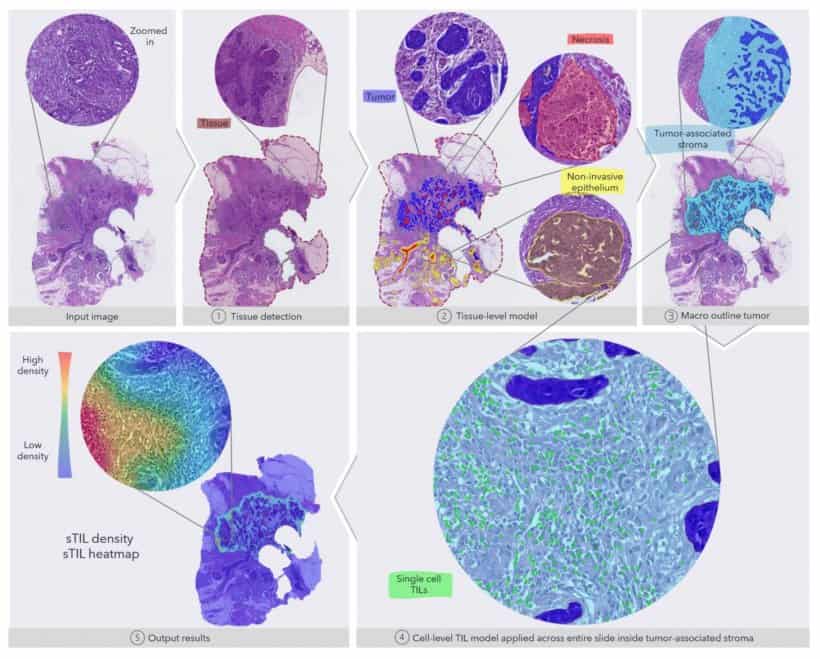
A researcher at the Technical University of Denmark (DTU) has developed a mathematical model for use in automated image analysis of tissue samples. The model provides the possibility for better and more similar cancer prognosis and treatment.

Algorithm detects prognostic immune signature in aggressive breast cancer using H&E images2021-07-16T07:42:01+00:00

A smartphone pathology platform to characterize tumor cytology and HER2 expression in breast cancer

2021-07-14T12:50:24+00:00

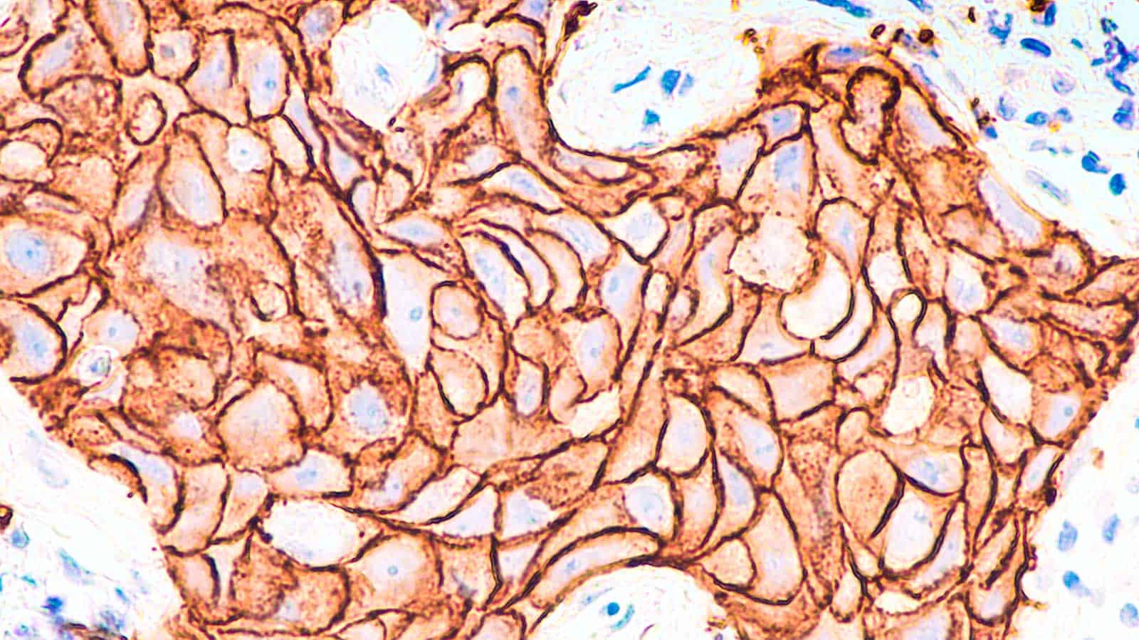
Scientists from the Department of Biomedical Engineering and the Department of Surgery, Duke University (Durham, NC, USA) have developed a multimodal mobile platform, EpiView-D4, as an economic diagnostic pathology approach for point-of-care characterization of the cellular morphology and molecular expression of clinically relevant biomarkers in fine-needle aspiration specimens from breast tumors.

A smartphone pathology platform to characterize tumor cytology and HER2 expression in breast cancer2021-07-14T12:50:24+00:00
Go to Top