Pathology News
admin

About admin@pathologynews.com

This author has not yet filled in any details.
So far admin@pathologynews.com has created 1089 blog entries.

September 2021

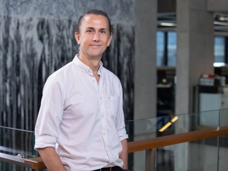
Conversation with Professor Gustavo Rohde – A Leading Expert in the Development of diagnostic AI Applications for Tissue Pathology.

2025-06-25T13:47:16+00:00

There is no doubt that exciting things are going to happen in the future, but it’s hard to predict which ones will be truly useful and have longevity. People in our field, the machine learners, they are also going to come up with innovations that will last. Some will be flashes in the pan, and some will continue and be pervasive and persistent in the future. If you ask me however, the solutions that are based on first discovering the fundamentals and then exploiting that knowledge to put together the algorithms, are the ones that will succeed, and that is where I tend to focus my time.

Conversation with Professor Gustavo Rohde – A Leading Expert in the Development of diagnostic AI Applications for Tissue Pathology.2025-06-25T13:47:16+00:00

Artificial intelligence predicts the risk of metastatic relapse in breast cancer patients

2021-09-22T02:34:57+00:00

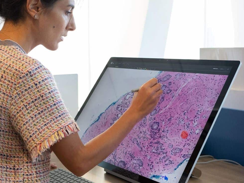
The RACE AI study conducted by Gustave Roussy and the startup Owkin, as part of the AI for Health Challenge organized by the Ile-de-France Region in 2019, was presented as a proffered paper at ESMO (European Society of Medical Oncology). This study shows that thanks to deep learning analysis applied to digitized pathology slides, artificial intelligence can classify patients with localized breast cancer between high risk and low risk of metastatic relapse in the next five years . This AI could thus become an aid to therapeutic decision making and avoid unnecessary chemotherapy and its impact on personal, professional and social lives for low risk women. This is one of the first proofs of concept illustrating the power of an AI model for identifying parameters associated with relapse that the human brain could not detect. With 59,000 new cases per year, breast cancer ranks first among cancers in women, clearly ahead of lung cancer and colorectal cancer. It is also the cancer that causes the greatest number of deaths in women, with 14% of female cancer deaths in 2018,. 80% of breast cancers are said to be hormone-sensitive or hormone-dependent. But these cancers are extremely heterogeneous and about 20% of patients will relapse with distant metastasis. RACE AI is a retrospective study that was conducted on a cohort of 1400 patients managed at Gustave-Roussy between 2005 and 2013 for localized hormone-sensitive (HR+, HER2-) breast cancer. These women were treated with surgery, radiotherapy, hormone therapy, and sometimes chemotherapy to reduce the risk of distant relapse. Chemotherapy is not routinely administered because not all women will benefit from it due to a naturally favorable prognosis. The practitioner's choice is based on clinico-pathological criteria (age of the patient, size and aggressiveness of the tumor, lymph node invasion, etc.) and the decision to administer or not adjuvant chemotherapy varies between oncology centers. Genomic signatures exist today to help identify women who benefit from chemotherapy, but they are not recommended by the French National Authority for Health and are not reimbursed by the French National Health Insurance (although they are included on the RIHN reimbursement list), which makes their access and use heterogeneous in France. Related StoriesGustave Roussy and Owkin have taken up the challenge of proposing a new method that is simple, inexpensive and easy to use in all oncology centers as a therapeutic decision-making tool. Ultimately, the goal is to direct patients identified as being at high risk towards new innovative therapies and to avoid unnecessary chemotherapy for low-risk patients. In the RACE AI study, Owkin's Data Scientists, guided by Gustave Roussy's research physicians, developed an AI model capable of reliably assessing the risk of relapse with an AUC of 81% to help the practitioner determine the benefit/risk balance of chemotherapy. This calculation is based on the patient's clinical data combined with the analysis of stained and digitized histological slides of the tumor. These slides, used daily in pathology departments by anatomo-pathologists, contain very rich and decisive information for the management of cancer. It is not necessary to develop a new technique or to equip a specific technical platform. The only essential equipment is a slide scanner, which is a common piece of equipment in laboratories. Like an office scanner that digitizes text, this scanner digitizes the morphological information present on the slide. The results of this first study by the Owkin and Gustave Roussy teams open up strong prospects and next steps include prospectively validating the model on an independent cohort of patients treated outside Gustave Roussy. If the results are confirmed, through providing reliable information to clinicians, this AI tool will prove to be a valuable aid to therapeutic decisions. Owkin, Inc

Artificial intelligence predicts the risk of metastatic relapse in breast cancer patients2021-09-22T02:34:57+00:00

Using deep learning to predict biomarker status of breast tumors

2021-09-20T09:53:32+00:00

Artificial intelligence (AI) and machine learning entail the use of machines and algorithms to carry out complicated tasks and solve complex problems. Deep learning is a machine learning method that involves the generation and implementation of artificial neural networks with three or more layers. By simulating human intelligence, these neural networks can “learn” from large amounts of [...]

Using deep learning to predict biomarker status of breast tumors2021-09-20T09:53:32+00:00

Breast cancer ‘ecotypes’ defined by cellular & spatial genomics technologies could lead to more personalised treatment

2025-11-24T16:43:46+00:00

A team led by the Garvan Institute of Medical Research has revealed a new approach for classifying breast cancer subtypes based on their cell profile, which could help personalise treatments for patients.

Breast cancer ‘ecotypes’ defined by cellular & spatial genomics technologies could lead to more personalised treatment2025-11-24T16:43:46+00:00

New method enables 3D imaging of human organs

2021-09-13T17:57:04+00:00

Researchers at Umeå University now demonstrate a method by which specific cell types in human organs can be studied with micrometer precision. The method can be used to reveal previously unrecognised alterations in the pancreas, but it can also be used to study other human organs and diseases.

New method enables 3D imaging of human organs2021-09-13T17:57:04+00:00

Researchers create detailed cell atlas of gut and reveal developmental origins of Crohn’s disease

2025-11-24T14:58:32+00:00

High magnification micrograph of Crohn's disease. Biopsy of esophagus. H&E stain. Credit: Nephron/Wikipedia Many diseases have their origin in early human development, and today (8 September), two publications in Nature reveal how researchers from the Human Cell Atlas (HCA) consortium are advancing understanding of this. The global HCA initiative is mapping every cell type in the human body, to transform our knowledge of biology, infection and disease. A new large-scale study mapped the cells in the human gut from early development through to adulthood, creating the most comprehensive cell atlas of the gut to date. This revealed that Crohn's disease may be caused by activation of developmental pathways, and uncovered potential drug targets for treating Crohn's and other inflammatory bowel diseases. The detailed maps will help explain how the gut forms and functions, and will transform research into intestinal diseases. The second publication reveals the hugely ambitious plan to create an entire human developmental cell atlas (HDCA) of all cells that are important for healthy human development. The gut is just one example of the importance of this, and researchers from the Human Cell Atlas Developmental Biological Network and their collaborators worldwide show how they will chart developing tissues comprehensively in space and time. Key to understanding what happens in early development and how this can affect health or lead to disease, the HDCA is likely to lead to transformations in healthcare. The gut is a complicated tissue made of multiple cell types, and changes enormously during early development. To understand how the gut develops and functions, researchers from the Wellcome Sanger Institute, Newcastle University, University of Cambridge and their collaborators within the Human Cell Atlas studied more than a third of a million individual gut cells from developing tissue and from child and adult donors. Using cutting edge single-cell genomics and spatial analysis techniques, the team revealed which genes were active in each cell, and created a highly extensive gut cell atlas through time and across 12 regions of the intestines. Rasa Elmentaite, first author on the study from the Wellcome Sanger Institute, said, "By studying multiple regions of the human gut throughout development, childhood and adulthood we've created a unique, detailed map of the healthy human gut. This gut cell atlas reveals complex developmental events, including how the immune and nervous systems develop in the healthy gut, and identifies important differences along the intestines. The data is openly available to other researchers studying the gut, and will undoubtedly contribute to future discoveries."

Researchers create detailed cell atlas of gut and reveal developmental origins of Crohn’s disease2025-11-24T14:58:32+00:00

Machine learning accelerates super-resolution microscopy

2021-09-09T14:17:01+00:00

The DECODE (DEep COntext DEpendent) algorithm is based on deep learning, but instead of using real images the network is trained with synthetic data generated by a numerical simulation. By incorporating information about the microscopic setup and the imaging physics, the researchers achieved simulations that closely matched real-world acquisitions.

Machine learning accelerates super-resolution microscopy2021-09-09T14:17:01+00:00

Olympus and Grundium join forces to advance digital pathology

2021-09-08T14:27:04+00:00

Olympus and Grundium have joined forces to enhance the implementation of digital pathology technologies in diagnostic and research laboratories. Olympus Corp. (OTCMKTS: OCPNY) is a leading manufacturer of optical and digital precision technology, designing and manufacturing microscopes for over 100 years. Olympus’ revolutionary products include X Line objectives, which are developed using a proprietary manufacturing technology. Grundium, [...]

Olympus and Grundium join forces to advance digital pathology2021-09-08T14:27:04+00:00

Paige’s AI Diagnostic Tech Is Revolutionizing Cancer Diagnosis

2021-09-03T13:22:46+00:00

The startup’s software analyzes tissue-sample imagesArtificial intelligence was not on Leo Grady‘s mind when he was applying for college in the early 1990s. The IEEE member was interested in human interaction, linguistics, and psychology, and he initially chose the unusual double major of anthropology and physics.But his desire to understand how the human brain works led him to switch from physics to electrical engineering and ultimately to pursue signal processing at the University of Vermont.“All of this eventually led me to AI,” Grady says. “AI is a mix of psychology, engineering, computer science, and a little bit of philosophy. It really caught my imagination.”Grady is now the chief executive of Paige, in New York City. In 2018 the company spun off from the Memorial Sloan Kettering Cancer Center. Paige uses machine learning to help pathologists make faster, more accurate cancer diagnoses from images of tissue samples. The startup, which is focusing on detecting prostate and breast cancer, plans to expand to other diseases.Paige hopes to help doctors catch cancer earlier, come up with effective treatment plans, and prevent unnecessary surgeries due to false positives. In May the company announced a partnership with Quest Diagnostics to develop software products that could find markers of cancer that might not have been known before, and that could help pathologists and oncologists better diagnose prostate, breast, and other types of cancer.“Our technology is truly transformative,” Grady says. “It’s going to help pathologists be more efficient, make higher quality decisions, and get faster results back to patients. It will also ultimately be less expensive for the health care system.”In January the startup raised US $100 million in Series C funding. Paige’s prostate cancer diagnostic technology is the first AI product designed for pathology or oncology to earn U.S. Food and Drug Administration (FDA) Breakthrough Device designation.Paige is now seeking to use its technology beyond diagnosis. By training its AI to understand the correlations between certain types of tumors and the effectiveness of certain drugs, Grady says, the company hopes to be able to predict treatment regimens for patients.HAVING A MEANINGFUL IMPACTPaige’s work is a continuation of the ambition Grady has had since he was a graduate student at Boston University—of making a difference in the world with AI. His Ph.D. thesis focused on the application of neural networks to image analysis. Neural networks are an approach to machine learning, loosely modeled on the human brain, that can be used to identify patterns in data sets. At the heart of today’s AI, they are used for Web searches, object detection, and facial recognition. In the early 2000s, though, neural network research “was on the fringe of AI, the black sheep,” Grady says.But Grady, who was finishing his doctorate around that time, saw the technology’s potential for the real world. He says he knew he did not want to pursue a career in academia: “I felt that publishing papers wasn’t enough.”To find a job where he could have a meaningful impact, Grady browsed issues of the IEEE Transactions on Pattern Analysis and Machine Intelligence, looking for research articles authored by experts in industry.“I saw name after name coming from Siemens,” he says, “so that was the place I wanted to go.”Siemens Corporate Technology, in Princeton, N.J., had one of the world’s best computer vision research groups at the time, he says. He joined in 2003 as a research scientist, developing computer vision software for the company’s imaging machines. He focused on medical image analysis, extracting pertinent information from scans of cardiovascular and cancer patients that could help with diagnosis. He was granted 40 U.S. patents in his almost nine years with the company.But despite the success of those AI-based medical analysis instruments in in-house tests, he “kept hitting roadblock after roadblock” when trying to introduce the machines into medical offices.“They weren’t getting used by doctors,” he says. “They’d say, ‘It doesn’t fit into the hospital’s IT system’ or ‘I can’t get paid for using them’ or ‘I don’t have time to do it.'”He soon realized that making inroads with AI software would be easier than selling a new hardware system, so he left in 2012 to join medical technology startup HeartFlow, in Redwood City, Calif., as vice president of research and development.There he led the development of a software-based diagnostic test for coronary heart disease. Starting with a cardiac CT scan, the technology used AI and fluid dynamics to build a 3D model of the heart, calculate blood flow, and help determine if a stent was needed. The approach allows doctors to avoid more-invasive tests, he says.“It’s better for patients and doctors,” he says, “because it’s lower-cost and lower-risk.”The technique, which has received clearance from the FDA, is now used by cardiologists in Europe, Japan, and the United States. HeartFlow in July announced plans to go public after merging with Longview Acquisition.GREAT OPPORTUNITYGrady joined Paige in 2019 because of the opportunity it offered him to impact the world with AI, he says, with products that could transform cancer care.Cancer pathology today involves examining tissue samples under a microscope to make diagnoses. But tissues and disease markers can vary widely, so it’s common for pathologists to seek a second opinion or conduct more tests.Paige’s technology streamlines the process by digitizing it. The company has exclusive rights to tens of thousands of already-analyzed pathology slides from Memorial Sloan Kettering and has scanned them into its system to create a database of high-resolution images. The company’s proprietary machine-learning system is trained to detect patterns in the images that correlate with disease prognosis.When a new tissue scan comes in, the system can classify it based on its training. Rather than phoning a colleague or doing extra testing, a pathologist can use the AI-based system to make decisions more quickly and easily, Grady says.He and his colleagues recently published results showing that Paige’s prostate test reduced diagnostic time by about 65 percent, and it identified prostate cancer in four patients whose cancers were not initially diagnosed by three experienced histopathologists.EXCELLENT JOURNALSGrady says he is “a big fan” of IEEE. He joined as an undergraduate student and visited the organization’s New Jersey headquarters when he worked at Siemens. The organization has played an important role in his path from engineering student to CEO, he says.The IEEE Transactions on Pattern Analysis and Machine Intelligence, Medical Imaging, and Image Processing “stand out as the highest excellence in technical journals in these spaces,” he says.The IEEE Computer Vision and Pattern Recognition conference, he says, was one of the earliest in its field and “the only venue to showcase such research for a long time.”“The 2003 conference is where I met leaders in the field,” he says. “Without those conferences and those publications, the field wouldn’t be what it is today.”His message to IEEE’s graduate student members is that joining a company doesn’t have to mean stifling your research interests.“People often say you’re either in research or in industry, selling out,” he says. “I think it’s a false choice. Work that’s changing the world is happening in industry.”FROM [IEEE] ARTICLESRELATED ARTICLES AROUND THE WEBSource: IEEE Spectrum

Paige’s AI Diagnostic Tech Is Revolutionizing Cancer Diagnosis2021-09-03T13:22:46+00:00
Go to Top