Validating Whole Slide Imaging Systems for Diagnosis: Updated Guidelines
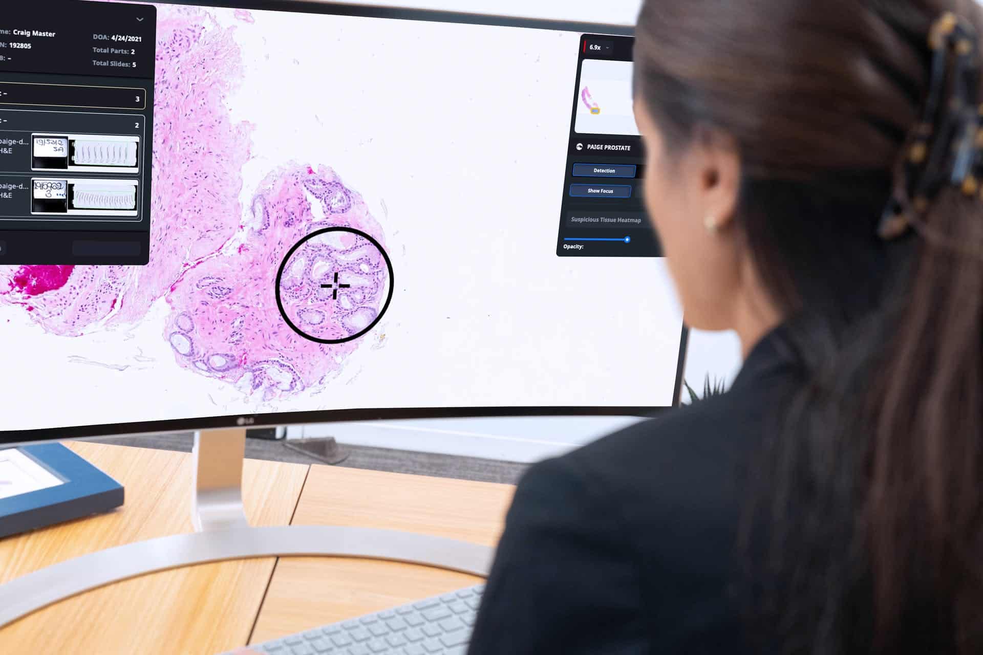
admin@pathologynews.com2022-01-04T13:14:39+00:00Whole slide imaging (WSI) entails the use of systems to scan entire glass slides to generate digital tissue images. Since the emergence of whole slide scanning systems approximately two decades ago, there have been significant advances in WSI technologies.1 The potentials of WSI in pathology and disease diagnosis are tremendous. Indeed, WSI is set to transform modern pathology and the way diseases are diagnosed based on tissue biopsies.2 Instead of observing stained tissue slides under microscopes used in traditional pathology, digital whole slide images can be reviewed using computerized systems. In addition to remote slide reviewing, WSI also facilitates easy sharing of pathology images among collaborators across the globe. The adoption of WSI in pathology laboratories has reached unprecedented levels in recent years, and despite its many advantages, guidelines are required to confirm the performance and diagnostic accuracy of WSI systems before they are used for diagnostic purposes.