Pathology News
admin

About admin@pathologynews.com

This author has not yet filled in any details.
So far admin@pathologynews.com has created 1089 blog entries.

April 2021

MUSE microscopy: using UV light to make histology rapid and slide-free

2021-04-20T00:03:56+00:00

Despite rapid technological advances in molecular diagnostics, optical microscopy remains the gold standard in pathology, cancer diagnosis, and disease monitoring. Considering that the optical principles of light microscopy and histological preparation methods have seen little change over decades or even centuries, the development of new microscopy and histological preparation methods seems to be long overdue. Limitations of [...]

MUSE microscopy: using UV light to make histology rapid and slide-free2021-04-20T00:03:56+00:00

Multiplex Imaging Reveals CD40-stimulating Drug Helps to Jumpstart a T-cell Attack in Early-stage Pancreatic Cancer

2025-11-24T15:53:05+00:00

Giving early-stage pancreatic cancer patients a CD40 immune-stimulating drug helped jumpstart a T cell attack to the notoriously stubborn tumor microenvironment before surgery and other treatments, according to a new study from researchers in the Abramson Cancer Center (ACC) at the University of Pennsylvania. Changing the microenvironment from so-called T cell "poor" to T cell "rich" with a CD40 agonist earlier could help slow eventual progression of the disease and prevent cancer from spreading in more patients.
Multiplex Imaging Reveals CD40-stimulating Drug Helps to Jumpstart a T-cell Attack in Early-stage Pancreatic Cancer2025-11-24T15:53:05+00:00

Configuring infrared spectroscopy tools to better detect breast cancer

2021-04-15T06:49:52+00:00

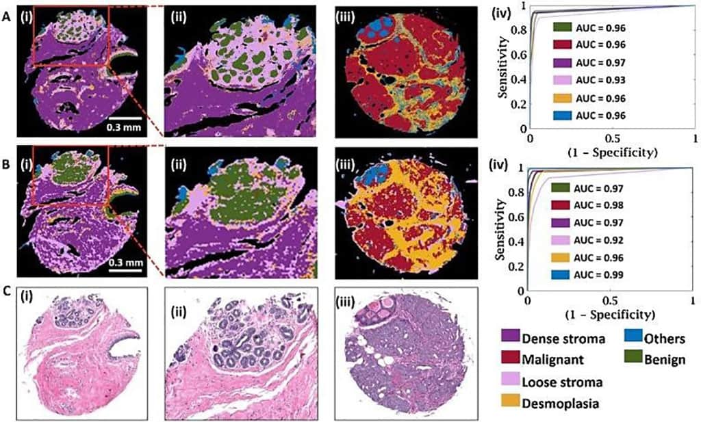
Digital analysis of cancer specimens using spectroscopic imaging coupled to machine learning is an emerging area that links spatially localized spectral signatures to tissue structure and disease. Breast histopathology, as an example of the broad relevance of these techniques, is critically important for clinical diagnoses. Current histologic characterization is morphology-based; thin tissue sections are stained, and cells are visually recognized by a pathologist using an optical microscope. However, the basis of the disease is well known to be molecular.

Configuring infrared spectroscopy tools to better detect breast cancer2021-04-15T06:49:52+00:00

Cooperation to accelerate adoption of AI-powered digital pathology

2021-04-09T07:09:01+00:00

Ibex’s Galen platform adds AI-powered cancer detection, case prioritization, grading and other productivity-enhancing insights. Users have reported significant improvements in diagnostic efficiency, with 27% reduction in time-to-diagnosis compared to conventional microscope viewing, 1- to 2-day reductions in total turnaround time, and 37% productivity gain3. In addition to cancer, the AI platform supports pathologists in the accurate grading, as well as detection and diagnosis of multiple clinical features, such as tumor size, perineural invasion, high-grade PIN (Prostatic Intraepithelial Neoplasia) and more.

Cooperation to accelerate adoption of AI-powered digital pathology2021-04-09T07:09:01+00:00

Artificial Intelligence: Harnessing the Full Potential of Digital Pathology in Routine Diagnosis

2021-04-09T00:01:20+00:00

  Artificial intelligence (AI) involves the use of machines and algorithms that have the ability to “think” or “act” as if they possessed human intelligence and critical thinking. Machine learning is a type of AI entailing computer software and algorithms that can learn from new data. “Trained” machine learning algorithms can make predictions, execute tasks, make data-driven [...]

Artificial Intelligence: Harnessing the Full Potential of Digital Pathology in Routine Diagnosis2021-04-09T00:01:20+00:00

Scientific repositories go digital

2021-04-08T23:57:08+00:00

Biobanks can be defined as a structured repository of biological specimens obtained from patients having a particular disease. These collections then serve as materials for future scientific and medical research, specifically, in translational medicine for the discovery of biomarkers and molecular factors involved in disease diagnosis, progression, and treatment. Biospecimens can be any many forms, such as [...]

Scientific repositories go digital2021-04-08T23:57:08+00:00

Experiences of Digitisation in HEp-2 Pattern Recognition – from Human to Machine

2021-04-08T12:27:24+00:00

A Brief Overview Human epithelial (HEp-2) cells are a vital tool in the diagnosis of autoimmune diseases. In recent years, there have been advancements to improve standardisation and quality of autoimmune diagnosis using indirect immunofluorescence assay (IIFA) on HEp-2 cells.   Many laboratories have switched over to using automated systems, that not only perform the assays but [...]

Experiences of Digitisation in HEp-2 Pattern Recognition – from Human to Machine2021-04-08T12:27:24+00:00

March 2021

Covid-19 mapping reveals organ distribution and tissue damage

2021-03-30T13:40:01+00:00

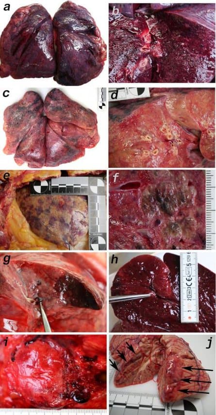
Macromorphology findings of 11 Covid-19 patients reveals involvement of multiple organs. (a) Pneumonectomy of patient one showed strong congestion with liquids and hemorrhages. The tissue consistency was fragile. (b) Cut surface of lung tissue in higher magnification as shown in (a). The pleura shows further hemorrhages. (c) Pneumonectomy of patient seven showed a more solid lung tissue without congestion. The tissue consistency was very firm. (d) Cut surface of lung tissue in higher magnification as shown in (c). Lung tissue was retracted adjacent to the bronchus. (e) Pale pleura visceralis of the lung of patient 6 with disseminated hemorrhages and signs of disturbed ventilation. (f) Nodular transformation of lung tissue as phenomenon of fungal superinfection in patient 3. (g) Hemorrhagic lung infarct in patient 4 due to a thrombembolus in a pulmonary artery branch. (h) Anemic spleen infarct due to a clotted small artery in patient 4. (i) Fulminant stasis and thromboses in the periprostatic plexus in patient 4. (j) Cerebellar infarction (hemorrhagic) in patient 9.Image source: Deinhardt-Emmer et al., eLife 2021 (CC BY 4.0) “Clinical observations suggest that Covid-19 is a systemic disease, meaning that it affects the entire body rather than just a single organ such as the lungs,” explains co-first author Stefanie Deinhardt-Emmer, Resident in Medical Microbiology, Jena University Hospital, Jena, Germany. “But we don’t currently have a clear understanding of disease development in humans and other organisms, due to the lack of appropriate experimental models. Investigating the viral distribution of SARS-CoV-2 within the human body and how this relates to tissue damage would help us address this gap.” To do this, Deinhardt-Emmer and colleagues studied 11 autopsy cases of patients with Covid-19. They performed the autopsies at the early postmortem stage to minimise bias due to the degradation of tissues and viral ribonucleic acid (RNA – a molecule similar to DNA).Their analysis revealed high viral loads in most of the patients’ lungs, which had caused significant damage to those organs. Using an imaging technique called transmission electron microscopy, the team also visualised intact viral particles in the lung tissue. “Interestingly, we also detected SARS-CoV-2 RNA throughout various other tissues and organs unrelated to the lungs that did not cause visible tissue damage,” says co-first author Daniel Wittschieber, Senior Forensic Pathologist at Jena University Hospital. The researchers say that this distribution of viral RNA throughout the body supports the idea that our immune system is unable to respond adequately to the virus’ presence in the blood. “We show that Covid-19 is a systemic disease as determined by the presence of virus RNA, and yet unrelated to tissue damage outside the lungs,” says co-senior author Bettina Löffler, Director of the Institute for Medical Microbiology, Jena University Hospital. “To our knowledge, this study is the only one to date that has measured viral loads in a wide variety of organs and tissues, with more than 60 samples studied per patient.”“The insights gathered from our work may add to our understanding of how Covid-19 develops in the body following infection,” concludes co-senior author Gita Mall, Head of the Institute of Forensic Medicine, Jena University Hospital.Source: Jena University Hospital

Covid-19 mapping reveals organ distribution and tissue damage2021-03-30T13:40:01+00:00

New Technique Provides Detailed Map of Lung Pathology in COVID-19

2025-11-24T16:10:18+00:00

A team led by investigators at Weill Cornell Medicine and NewYork-Presbyterian has used advanced technology and analytics to map, at single-cell resolution, the cellular landscape of diseased lung tissue in severe COVID-19 and other infectious lung diseases. In the study, published online March 29 in Nature, the researchers imaged autopsied lung tissue in a way that simultaneously highlighted dozens of molecular markers on cells. Analyzing these data using novel analytical tools revealed new insights into the causes of damage in these lung illnesses and a rich data resource for further research. "COVID-19 is a complex disease, and we still don't understand exactly what it does to a lot of organs, but with this study we were able to develop a much clearer understanding of its effects on the lungs," said co-senior author Dr. Olivier Elemento, professor of physiology and biophysics, director of the Caryl and Israel Englander Institute for Precision Medicine, associate director of the HRH Prince Alwaleed Bin Talal Bin Abdulaziz Alsaud Institute for Computational Biomedicine at Weill Cornell Medicine and co-Director of the WorldQuant Initiative for Quantitative Prediction, which funded the technology for single cell analysis of tissue. "I think the technological approach we used here is going to become standard for studying such diseases." Traditional tissue analysis, often using chemical stains or tagged antibodies that label different molecules on cells and can reveal important features of autopsied tissues. However, this approach is limited in the number of features it can mark simultaneously. It also usually doesn't allow detailed analyses of individual cells in tissues while retaining information about where the cells were in the tissue. The main technology the investigators employed in the study, a technology called imaging mass cytometry, largely overcomes those limitations. It uses a collection of metal-tagged antibodies that can simultaneously label up to several dozen molecular markers on cells within tissues. A special laser scans the labeled tissue sections, vaporizing the metal tags, and the metals' distinct signatures are detected and correlated with the laser position. The technique essentially maps precisely where cells are in the sample as well as each cell's surface receptors and other important identifying markers. Altogether over 650,000 cells were analyzed. The researchers applied the method to 19 lung tissue samples autopsied from patients who had died of severe COVID-19, acute bacterial pneumonia, or bacterial or influenza-related acute respiratory distress syndrome, plus four lung tissue samples autopsied from people who had had no lung disease.

New Technique Provides Detailed Map of Lung Pathology in COVID-192025-11-24T16:10:18+00:00
Go to Top