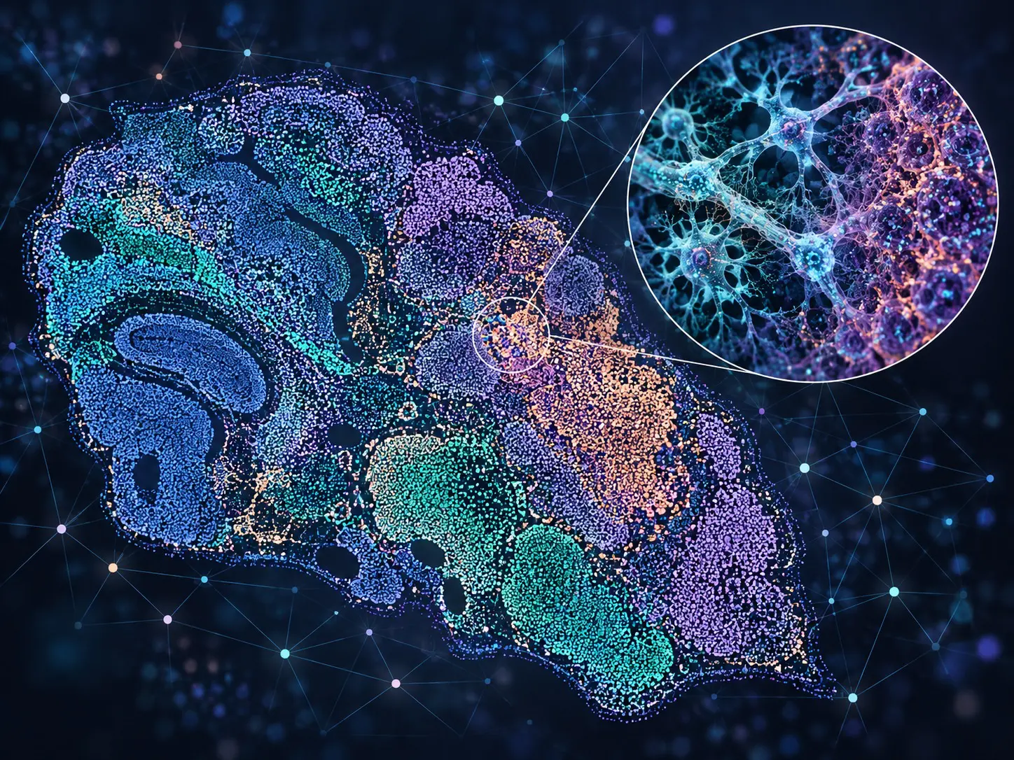
The following abstract is drawn from a recently published paper in Journal of Pathology Informatics | ScienceDirect.com by Elsevier. We invite you to read the full paper and join the conversation, become a member of the Pathology News community to share your thoughts, ask questions, and engage with others around this work.
Authors: Michail Georgios Papachristosa, Emiliano Spezib, Carolina Fuentesc, Rachel J. Erringtona, Dimitris Parthimosa
Abstract
Prostate cancer progression is routinely quantified by expert pathologists on hematoxylin and eosin (H&E)-stained tissue using Gleason grading. However, tissue processing for H&E staining is very time-consuming, whereas Gleason grading is often subject to inter- and intraobserver variability. This diagnostic variability can be reduced through utilization of mathematically quantitative and standardized metrics to analyze the prostatic tissue microenvironment. In this study, we identify an optimal set of metrics that could potentially be used as a diagnostic biomarker signature, through a combination of tissue architectures and gradient boosting algorithms. We employed fluorescence microscopy imaged prostatic tissue, stained with DRAQ5&Eosin, which allows for rapid real-time staining and imaging of the tissue, as opposed to H&E, and also provides high-resolution nuclear and cytoplasmic images. We then used engineered algorithmic pipelines to extract intensity-based, morphological, and textural features from four different tissue components: epithelial nuclei, epithelial cytoplasm, stromal nuclei, and stromal cytoplasm. We developed classification models to distinguish healthy from cancerous prostatic regions based on the extracted features and identified the most predictive features. Our epithelial-based models achieved area under the curves (AUCs) in the range 0.646–0.896, whereas our stromal-based models attained AUCs in the range 0.742–0.840. Our DRAQ5&Eosin-based models were thus able to identify important tissue specific spatial biomarkers, allowing for the creation of nuclear and cytoplasmic spatial profiles for healthy and cancerous prostatic tissue respectively.
Read the full article: Spatial characterization of prostate cancer microenvironment with DRAQ5&Eosin fluorescence imaging and quantitative feature identification – ScienceDirect
- aSchool of Medicine, Division of Cancer and Genetics, Cardiff University, UHW Main Building, Heath Park, Cardiff CF14 4XN, UK
- bDepartment of Biomedical Engineering & Biotechnology, Khalifa University, Shakhbout Bin Sultan St, Hadbat Al Za‘Faranah, Zone 1, Abu Dhabi, United Arab Emirates
- cSchool of Computer Science and Informatics, Cardiff University, Abacws, Senghennydd Road, Cardiff CF24 4AG, UK