Pathology News
admin

About admin@pathologynews.com

This author has not yet filled in any details.
So far admin@pathologynews.com has created 1089 blog entries.

April 2025

Indica Labs Announces Launch of High Dimensional Analysis Module for the HALO® Image Analysis Platform

2025-04-09T10:26:43+00:00

Indica Labs today announces the launch of the High Dimensional Analysis module for the HALO® image analysis platform. This exciting new functionality for customers in the life sciences enables advanced phenotypic characterization and spatial analysis as well as the exploration of highly multiplexed datasets in either brightfield or fluorescence.

Indica Labs Announces Launch of High Dimensional Analysis Module for the HALO® Image Analysis Platform2025-04-09T10:26:43+00:00

Transforming Prostate Biopsy Workflows with AI-Powered Digital Pathology

2025-04-08T10:46:08+00:00

Traditional pathology workflows, while effective, have inherent inefficiencies and limitations—especially for high-volume samples like prostate biopsies. Over decades, the promises of digital pathology often failed to deliver significant improvements. However, with the advent of Lumea’s technologies—like the BxBoard® and BxChip®—the field is finally witnessing measurable advances in efficiency, diagnostic accuracy, and patient outcomes.

Transforming Prostate Biopsy Workflows with AI-Powered Digital Pathology2025-04-08T10:46:08+00:00

Conversation with Prasanth Perugupalli, Pramana

2025-08-12T13:35:19+00:00

Join us for this Thought Leaders episode as Prasanth Perugupalli, Chief Product Officer at Pramana, shares his insights on the evolution of computational pathology, the role of AI in unlocking data from whole slide images, and what it takes to drive real-world adoption in labs around the world.

Conversation with Prasanth Perugupalli, Pramana2025-08-12T13:35:19+00:00

From Diagnosing to Disrupting: A Pathologist’s Leap into Innovation

2025-04-04T15:18:23+00:00

In this episode, Dr. Matthew O. Leavitt takes us through his digital pathology journey and what he’s learned along the way. Dr. Leavitt’s vision has always been to use digital technologies to improve care, especially in remote areas. He shares his experiences implementing digital pathology, the challenges faced, and the realization that standardization of tissue handling (pre-analytics) is crucial for efficient digital pathology workflows.

From Diagnosing to Disrupting: A Pathologist’s Leap into Innovation2025-04-04T15:18:23+00:00

Cheers to USCAP 2025: Recapping The Momentum, Insights, and Inspiration

2025-04-04T10:31:03+00:00

The United States and Canadian Academy of Pathology (USCAP) Annual Meeting is always a standout—a forum for trading the latest insights, engaging with the Concentriq community, and enjoying time with our team. This year’s event was no exception. Digital pathology and AI took center stage as the focus on precision medicine was stronger than ever. We left [...]

Cheers to USCAP 2025: Recapping The Momentum, Insights, and Inspiration2025-04-04T10:31:03+00:00

U.S. FDA Grants Paige Breakthrough Device Designation for AI Application that Detects Cancer Across Different Anatomic Sites

2025-04-04T09:00:50+00:00

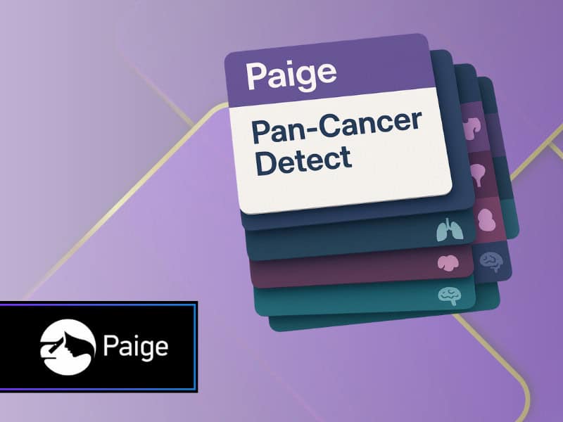
Paige, a leader in next-generation AI technology, today announced that the U.S. Food and Drug Administration (FDA) has granted Breakthrough Device designation to Paige PanCancer Detect*, an AI-assisted diagnostic application intended to assist pathologists in the detection of foci that are suspicious for cancer across a wide range of tissue from multiple organs.

U.S. FDA Grants Paige Breakthrough Device Designation for AI Application that Detects Cancer Across Different Anatomic Sites2025-04-04T09:00:50+00:00

ALAFIA Sets the Bar for Digital Pathology Automated Whole Slide Image (WSI) Processing in Under 35 Seconds

2025-05-28T14:50:01+00:00

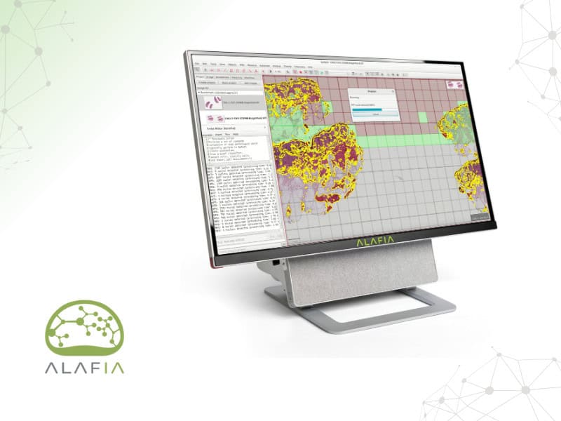
Traditionally, pathologists diagnose cancer and rare diseases by looking for abnormalities in tumor tissue and cells under a microscope, however, that is a time-consuming process often prone to errors. With the advent of whole slide imaging, pathologists are slowly migrating to a fully digital workflow. Whole slide imaging is the process of digitization of tissue samples on glass slides using digital whole slide scanners, enabling clinicians in histopathology, immunohistochemistry, and cytology to view, manipulate, interpret and digitally store the digital images, optimizing their workflow. Digitization also allows pathologists to interpret images using computational approaches, with the potential to improve accuracy, reduce inter-observer variability and provide new insights from a patient’s biopsy. Ultimately digital and computational pathology workflows will help clinicians and researchers discover, diagnose and treat diseases like cancer faster.

ALAFIA Sets the Bar for Digital Pathology Automated Whole Slide Image (WSI) Processing in Under 35 Seconds2025-05-28T14:50:01+00:00

Diagnexia and Stratipath Link Up Brings AI Breast Cancer Diagnostics to UK

2025-04-03T10:38:47+00:00

Diagnexia has announced a partnership with Stratipath AB to bring Stratipath Breast, an AI-based prognostic risk profiling tool, to healthcare providers across the UK and Europe.

Diagnexia and Stratipath Link Up Brings AI Breast Cancer Diagnostics to UK2025-04-03T10:38:47+00:00

Deep Bio to Present Cutting-Edge AI-Powered Research in Digital Pathology at AACR 2025

2025-04-02T13:32:47+00:00

Deep Bio, a pioneer in AI-powered digital pathology, will present new research at the American Association for Cancer Research (AACR) Annual Meeting 2025, highlighting the role of artificial intelligence in enhancing biomarker quantification and cancer diagnostics. The company’s findings will be showcased in multiple poster presentations based on deep learning models for PD-L1 and c-MET IHC quantification and frozen-section lymph node diagnosis.

Deep Bio to Present Cutting-Edge AI-Powered Research in Digital Pathology at AACR 20252025-04-02T13:32:47+00:00

AIVIS Announces MFDS Approval for AI-Driven Ki-67 Quantification Solution Using Microscope-Captured Images

2026-01-19T09:23:36+00:00

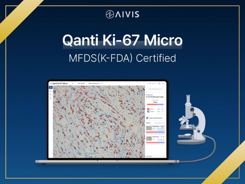
AIVIS, a leading medical AI company specializing in digital pathology, announced that it has received regulatory approval from South Korea’s Ministry of Food and Drug Safety (MFDS) for its latest AI-powered in vitro diagnostic software, Qanti Ki-67 Micro. The newly approved solution enables automated quantification of Ki-67—a key biomarker used to assess tumor cell proliferation—using only microscope-captured images, reducing reliance on capital-intensive digital scanners and offering a more accessible alternative.

AIVIS Announces MFDS Approval for AI-Driven Ki-67 Quantification Solution Using Microscope-Captured Images2026-01-19T09:23:36+00:00
Go to Top