by Christos Evangelou, MSc, PhD – Medical Writer and Editor
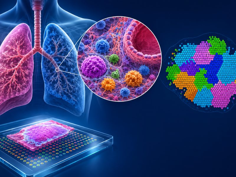
A recent spatial biology analysis demonstrated that the airways of individuals with asthma and chronic obstructive pulmonary disease (COPD) have distinct cellular neighborhoods. Using spatial profiling, researchers at the Mayo Clinic found that CD8+ T cells play a more prominent role in asthma than previously recognized, while COPD airways exhibit characteristic matrix-enriched niches that could guide the selection of anti-fibrotic therapies.
The study was published in Respiratory Research.
Study Rationale
Research using isolated cells falls short in revealing the role of the tissue context and spatial cell relationships in respiratory diseases, explained Y.S. Prakash, MD, PhD, senior author and Professor of Anesthesiology and Physiology at Mayo Clinic.
“We knew that asthma and COPD involve intricate crosstalk between multiple cell types, including epithelium, smooth muscle, fibroblasts, and immune cells, but we couldn’t see where and how these interactions occur in their native environment,” Prakash stated.
Methodology
The research team used the Akoya PhenoCycler-Fusion system to examine lung tissue samples from 17 individuals: five without asthma, four with asthma, and eight with COPD. They used a 10-marker antibody panel to map immune cells (CD45, CD3, CD4, CD8), structural components (E-cadherin, smooth muscle actin, collagen), and functional markers (Ki67, PCNA for proliferation, CD34 for angiogenesis) at single-cell resolution.
"Selecting our 10-marker panel was like trying to capture a symphony with just a few instruments," noted Latifa Khalfaoui, MD, PhD, the first author of the study. "We strategically chose markers that would allow us to distinguish major cell lineages and functional states while working within current technological constraints."
Formalin-fixed paraffin-embedded tissue samples were processed through multiple imaging cycles, with each cycle capturing three fluorescent markers plus nuclear staining. The team developed computational models using QuPath software and machine learning-based segmentation to identify and classify individual cells within their spatial context.
The analysis pipeline included unsupervised clustering to identify seven cell classes: epithelium, matrix cells, cytotoxic T cells, helper T cells, smooth muscle cells, other immune cells, and unclassified cells. Spatial analysis was used to examine cell-to-cell proximity patterns and identify recurring cellular neighborhoods within the tissue architecture.
Spatial Patterns in Lung Tissues
The study showed an increase in CD8+ T cells in samples from patients with asthma compared to those without asthma and patients with COPD. According to the authors, this finding challenges the traditional view of asthma as primarily driven by CD4+ Th2 cells.
"We were surprised by the prominence of CD8+ T cells in asthmatic samples," Khalfaoui stated. "This aligns with emerging research showing that CD8+ T cells and group 2 innate lymphoid cells may be particularly important in severe, steroid-resistant asthma."
Spatial analysis demonstrated that CD8+ T cells in lung tissues from patients with asthma are not distributed randomly but rather form clusters near the epithelium while avoiding matrix cells. This spatial signature suggests they may play a more active role in epithelial remodeling than previously appreciated. In contrast, COPD samples showed higher abundance of matrix cells (~40% of all detected cells) compared to samples from patients with asthma (~28%) and those from individuals without asthma (~32%).
Cellular Neighborhoods Define Disease Status
The research team identified seven distinct cellular neighborhoods or niches within lung tissue. Each niche represents a recurring pattern of cellular organization that appears to be disease specific.
Lung tissues from patients with asthma showed a high frequency of immune cell-enriched niches and unclassified cell-enriched neighborhoods, suggesting active inflammatory remodeling processes. In contrast, lung tissues from patients with COPD were enriched for collagen-heavy matrix niches, reflecting the fibrotic nature of the condition.
"The clinical reality is that patients with similar diagnoses can have vastly different responses to treatment, suggesting underlying heterogeneity that our traditional approaches weren't capturing," Prakash explained. "Spatial biology offered us the opportunity to map these 'cellular neighborhoods' and understand how the tissue microenvironment itself contributes to disease pathogenesis and potentially treatment resistance."
Potential Clinical Implications
According to the authors, spatial analysis of lung tissues could help clinicians move beyond traditional histologic grading to identify patients who might benefit from specific treatments. Patients with matrix-enriched signatures might respond better to anti-fibrotic therapies, while those with immune-enriched signatures could benefit from targeted immunomodulatory therapy.
"The distinct cellular neighborhoods we identified could serve as spatial biomarkers for disease endotyping and treatment selection," Khalfaoui said. "COPD patients showed enrichment in collagen-heavy matrix niches, while asthmatics had more immune cell-enriched neighborhoods."
Looking Ahead
The researchers acknowledge that the 10-marker panel resulted in a high proportion of unclassified cells (50% in asthmatic samples).
"Rather than seeing this as purely a limitation, it actually highlights the incredible cellular diversity in diseased airways that we're only beginning to understand," Khalfaoui noted.
Prakash revealed that the team is planning studies to expand the marker panel to characterize additional cell types, including regulatory T cells, different macrophage polarization states, detailed epithelial cell subtypes, and specific extracellular matrix components.
Prakash also predicted that the integration of spatial proteomics with spatial transcriptomics represents the next frontier in precision medicine.
"While our protein data shows us the functional 'end state' of cells, spatial transcriptomics could reveal the dynamic gene expression programs driving these cellular neighborhoods," he explained. "The combination would give us both the 'what', through protein expression, and the 'why', through gene regulation of disease pathogenesis in spatial context."
Prakash concluded by emphasizing that the integration of spatial proteomics with spatial transcriptomics may help clinicians understand the temporal dynamics of cell neighborhoods, how they form, evolve, and potentially resolve with treatment.
"This could fundamentally change how we approach precision medicine in respiratory diseases," he added.
The study received financial support from the National Institutes of Health (NIH). The investigators would like to extend our sincere appreciation to the Mayo Office of Core Shared Services (OCSS) for their invaluable support in making their study possible. Special thanks are also due to the Immuno–monitoring Core (IMC), Pathology Research Core (PRC), and Bioinformatics Core for their significant contributions.
References
- Khalfaoui L, Moore RM,Villasboas JC, et al. Spatial phenotyping of human bronchial airways in obstructive lung disease. Respir Res. 2025;26(1):232. Published 2025 Jul 2. doi:10.1186/s12931-025-03315-5
No audio available for this article yet.
No quiz available for this article yet.