by Christos Evangelou, MSc, PhD – Medical Writer and Editor
Researchers at the University of Modena and Reggio Emilia in Italy and the University of Helsinki in Finland have developed an AI framework called PATHOS (Pathology Attention Framework) that analyzes routine pathology slides to identify tissue features associated with response to neoadjuvant chemotherapy in patients with high-grade serous ovarian carcinoma. According to the authors, the system could potentially enable more accurate patient stratification and personalized therapy decisions.
The study was published in the Journal of Pathology Informatics.
Study Rationale
Ovarian high-grade serous carcinoma (HGSC) has a poor prognosis, with five-year survival rates of approximately 25% for patients with advanced disease receiving neoadjuvant chemotherapy (NACT) before debulking surgery. The considerable variation in how patients respond to platinum-based chemotherapy creates a need for better predictive tools.
"This study was motivated by a concrete clinical need expressed by pathologists: the lack of tools that clearly link specific morphological features in histology to patient outcomes after treatment," explained Marta Lovino, PhD, who is an assistant professor at the University of Modena and Reggio Emilia and the corresponding author of the study. "In high-grade serous ovarian carcinoma, understanding which tissue characteristics are associated with treatment response remains challenging, yet clinically crucial."
Current methods of predicting treatment response entail manual assessment of treatment-induced changes in tissue architecture by expert pathologists. However, these morphological alterations can be subtle and heterogeneous. Scoring systems such as the Chemotherapy Response Score quantify fibroinflammatory stromal content, but they do not capture the full spectrum of post-treatment morphological features that may provide prognostic information.
Methodology: A Three-Module Architecture
The research team developed PATHOS to process hematoxylin and eosin (H&E) stained whole-slide images through three integrated modules that work sequentially to maximize both predictive accuracy and clinical interpretability.
The first module employs Multiple Instance Learning (MIL) with attention mechanisms to identify the most informative regions within tissue slides, rather than analyzing the entire slides. The team selected BufferMIL, a model that identifies a batch of relevant patches rather than single points, to better capture the heterogeneous nature of ovarian HGSC. This module achieved 89% accuracy and an area under the curve (AUC) of 93% in predicting progression-free interval.
"We developed a computational framework that analyzes routine H&E histology images and highlights which morphological components of the tissue are most informative of treatment response," Lovino said. "The method helps connect what pathologists see under the microscope with how patients actually respond to therapy."
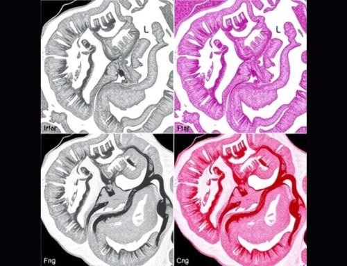
The second module performs panoptic segmentation, which identifies both tissue regions (stroma, tumor, necrosis, hemorrhage) and individual cell types (neoplastic, connective, inflammatory, dead cells, macrophages). The module uses a Cellpose-based model trained on manually annotated ovarian HGSC images and generates detailed spatial maps of tissue architecture. The analysis then extracts 69 morphological and spatial features, including tumor cell shape complexity, stromal abundance, and cell-cell proximity patterns.
The third module employs multiple machine learning classifiers to determine which features most strongly predict treatment response. The module uses model-agnostic interpretability methods, specifically SHAP (Shapley Additive Explanations) and LIME (Local Interpretable Model-agnostic Explanations), and identified seven key features that consistently distinguished between short and long platinum-free intervals.
Morphological Signatures of Treatment Response
The research team analyzed 176 H&E-stained whole-slide images from omental tumor tissue collected during interval debulking surgery from 50 patients with HGSC who were enrolled in the DECIDER clinical trial. Patients were stratified into two groups: those with progression-free intervals exceeding 365 days (indicating good platinum sensitivity) and those with intervals under 180 days (suggesting platinum resistance).
The seven features identified as most predictive of response showed higher values in patients with longer platinum-free intervals. These included maximum tumor cell eccentricity (degree of elongation), maximum tumor cell major axis length, mean tumor cell fractal dimension (structural complexity), number of stromal cells, distal stromal area, total stromal area, and stromal cell proportion. Patients with long progression-free intervals demonstrated increased tumor cell pleomorphism (irregular, complex cell shapes) and greater stromal content following chemotherapy than those with short progression-free intervals.
"The key message is that detailed morphological patterns in post-treatment tumor tissue carry meaningful information about patient response," Lovino noted. "Rather than relying on global or coarse features, our approach captures fine-grained tissue characteristics that are strongly associated with outcome. The consistency of these associations across patients was particularly encouraging."
Efficiency Through Selective Analysis
The MIL attention mechanism identified 9.77% of tissue patches as highly relevant for prediction, improving computational efficiency compared with analyzing entire slides.
The researchers compared classification using features extracted from attention-identified regions versus whole slides. XGBoost, the best-performing model on attention-guided features, demonstrated 92% accuracy and 98% AUC. The same model analyzing whole-slide features showed 60% accuracy and 58% AUC. HistGradientBoosting, the best performer on the entire slides, showed 99% AUC when applied to filtered patches, compared with 61% on whole slides. The framework also demonstrated discriminative ability for overall survival in Kaplan-Meier analysis, despite being trained on platinum-free interval.
Clinical Translation and Future Directions
"Clinically, these parameters could support more accurate stratification of treatment response in patients with ovarian cancer," Lovino said. "In the future, such information may help guide therapeutic decisions and improve personalized treatment strategies."
According to the authors, the morphological features identified using PATHOS may reflect underlying molecular heterogeneity in treatment response. The combination of attention maps showing which tissue regions drive predictions and quantitative features that pathologists can conceptually verify creates a framework that enables clinical experts to understand and validate algorithmic decisions.
"The most novel aspect lies in the level of morphological detail captured and interpreted in the specific context of high-grade serous ovarian carcinoma following neoadjuvant chemotherapy," Lovino explained. "The framework goes beyond prediction by offering interpretable links between tissue morphology and clinical outcome."
Study limitations include the relatively patient cohort (50 patients from a single clinical trial) and the fact that the framework focused specifically on omental metastases, which, while commonly sampled and prognostically important in HGSC, represent only one metastatic site.
"An important open question concerns large-scale validation," Lovino acknowledged. "Future studies are needed to assess the robustness and generalizability of these morphological parameters across larger and more diverse patient cohorts."
References
- Miccolis F, Lovino M, Lehtonen O, Hynninen J, Hautaniemi S, Virtanen A, Ficarra E. PATHOS: Pathology attention framework for treatment response stratification in ovarian high-grade serous carcinomas following neoadjuvant chemotherapy on H&E images. Journal of Pathology Informatics. 2026 Jan 21:100545.
No audio available for this article yet.
No quiz available for this article yet.