by Christos Evangelou, MSc, PhD – Medical Writer and Editor
A collaborative effort involving universities and research institutes in China and Germany led to the development of a deep learning model trained on standard hematoxylin and eosin (H&E)-stained whole-slide images to predict tumor lactate metabolic status, a feature that is typically assessed using molecular profiling. The researchers validated the model across 13 cancer types and in an independent real-world cohort, and found that it accurately predicted tumor lactate metabolism from routine histology, providing a scalable and practical digital biomarker for metabolism-informed precision oncology.
The study was published in Frontiers in Immunology.
Study Rationale
Lactate, the end product of aerobic glycolysis, acidifies the tumor microenvironment (TME), inhibits cytotoxic T cells and natural killer cells, promotes regulatory T cell expansion, and skews macrophage polarization toward an immunosuppressive M2-like state, explained Dr. Bohai Feng, one of the corresponding authors of the study. More recently, lactylation (post-translational modification of histones by lactate) has been identified as an additional mechanism through which elevated lactate reprograms both immune and stromal cells in the TME.
“Metabolic reprogramming is a fundamental feature of cancer, and lactate metabolism has emerged as an important regulator of tumor–immune interactions and therapeutic resistance,” Feng said. “However, assessing lactate metabolic activity in clinical samples usually necessitates molecular profiling, which is not always available in routine practice.”
The team aimed to explore whether tumor lactate metabolism could be inferred from standard pathology images using integrative multi-omics and digital pathology approaches.
Methodology
The research team constructed a 59-gene lactate-related signature, selecting protein-coding genes from the GeneCards database with a relevance score of 10 or higher. Protein–protein interaction (PPI) network analysis using STRING revealed five functional clusters within this gene set, namely respiratory electron transport, pyruvate metabolism, primary mitochondrial disease, lactate transmembrane transport, and aminoacyl-tRNA biosynthesis.
Using single-sample gene set enrichment analysis (ssGSEA), the team calculated lactate activity scores across multiple datasets. In a pre-treatment head and neck squamous cell carcinoma (HNSCC) dataset, non-responders to immune checkpoint inhibitors (ICIs) had higher lactate scores than responders. In two independent melanoma immunotherapy cohorts, patients in the high-lactate group had worse overall survival.
Among patients with HNSCC receiving adjuvant radiotherapy in the TCGA dataset, high lactate scores independently predicted worse overall survival, progression-free interval, and disease-specific survival. However, lactate scores did not predict outcomes in patients who did not receive radiotherapy.
Mapping the Immunosuppressive Microenvironment
The researchers classified TCGA-HNSCC samples into the top third (LAC_H) and bottom third (LAC_L) by lactate score and profiled the TME of each group using transcriptomic signatures. LAC_H tumors were enriched for MYC targets, oxidative phosphorylation, fatty acid metabolism, mTORC1 signaling, and DNA repair pathways. On the other hand, LAC_L tumors showed higher T cell, NK cell, and B cell infiltration, elevated MHC-I and co-stimulatory molecule expression, and stronger ICI-related ligand–receptor signaling.
“Our results show that tumors with high lactate metabolic activity exhibit increased proliferation and suppressed immune infiltration,” said Prof. Jochen Hess, one of the corresponding authors of the study. “These tumors also have poorer responses to immunotherapy and radiotherapy.”
In an additional single-cell RNA-seq analysis integrating data from 74 patients with HNSCC, malignant epithelial cells were enriched in LAC_H tumors, while fibroblasts, macrophages, and immune subsets were depleted. Twelve of the 59 lactate-related genes showed preferential expression in malignant cells. Histologically, LAC_H tumors appeared morphologically “cleaner” on H&E characterized by compact tumor nests with sparse immune infiltration. In contrast, LAC_L tumors displayed a more heterogeneous architecture with abundant immune and stromal components.
Teaching a Model to See Metabolism
“We integrated multi-omics analyses with spatial and pathological information to develop a deep learning model that can predict lactate metabolic states directly from routine, histologically stained, whole-slide images,” Hess noted.
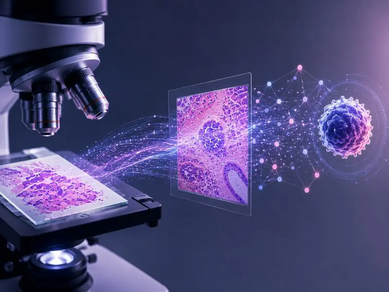
The computational pipeline entailed tiling digitized H&E slides into 512 × 512-pixel patches at 20× magnification, applying background exclusion and Vahadane color normalization, and extracting Patch-level features using the pre-trained CTransPath network. The process yielded 768-dimensional embeddings per patch, which were linearly projected to 512 dimensions and aggregated into slide-level representations through a two-layer transformer architecture with multi-head self-attention.
PCA compression to 128 dimensions and LASSO-based feature selection were applied before training four classifiers (XGBoost, Gradient Boosting, LightGBM, and SVM). In the TCGA-HNSCC test cohort, all four models distinguished LAC_H from LAC_L tumors with AUROCs ranging from 0.73 to 0.82.
Pan-Cancer Generalization and Protein-Level Validation
The team then extended the analysis to 12 additional TCGA solid tumor types, where the SVM model achieved AUROCs between 0.78 and 0.89.
“One particularly encouraging finding was the strong generalizability of the model across multiple cancer types,” Hess noted.
The team also applied their integrated lactate prediction score to 110 patients from the SAZHU-HNSCC cohort, an independent, real-world dataset of untreated surgical HNSCC specimens collected at Zhejiang University School of Medicine. The top and bottom 10% of predicted scores were subjected to quantitative immunohistochemistry for LDHA (the enzyme converting pyruvate to lactate) and MCT1 (the primary lactate transporter). Both markers showed higher H-scores in model-predicted high-lactate tumors. High-lactate cases displayed compact tumor nests with strong LDHA and MCT1 immunoreactivity, whereas low-lactate cases showed immune cell infiltration alongside diminished staining.
Clinical Implications and Next Steps
“This work demonstrates that images routinely collected for histopathological diagnosis may provide clinically meaningful information about tumor metabolism,” Feng said. “A digital biomarker that reflects lactate metabolic activity could help identify patients with an immunosuppressive tumor microenvironment and guide therapeutic strategies informed by metabolism.”
The authors acknowledge that the SAZHU-HNSCC cohort used for IHC validation included only 22 patients at the extremes of the predicted lactate distribution, and it originated from a single center. The integrated lactate prediction score was built on retrospective data, and whether it adds value to prospective clinical decision-making remains untested. The model also does not directly measure intratumoral lactate concentrations.
“Further studies are needed to investigate the mechanistic links between lactate metabolism and immune regulation,” Hess said. “The predictive value of the digital lactate biomarker must be evaluated prospectively in clinical trials and treatment decision-making.”
Feng concluded by emphasizing that the integration of multi-omics analysis with digital pathology bridges molecular metabolic states and tissue morphology, which could “enable direct inference of tumor metabolic activity from standard H&E slides and provide a scalable, cost-effective approach for clinical implementation.”
The study received financial support from the China Scholarship Council, the Deutsche Forschungsgemeinschaft, and the Zhejiang Provincial Natural Science Foundation of China.
References
- Feng B, Zhu Y, Zhang Z, Wang Y, Schuler PJ, Hess J. Tumor lactate metabolism shapes immune suppression and therapeutic resistance revealed by integrative multi-omics and digital pathology. Front Immunol. 2026;17:1797798. Published 2026 Mar 26. doi:10.3389/fimmu.2026.1797798