by Christos Evangelou, MSc, PhD – Medical Writer and Editor
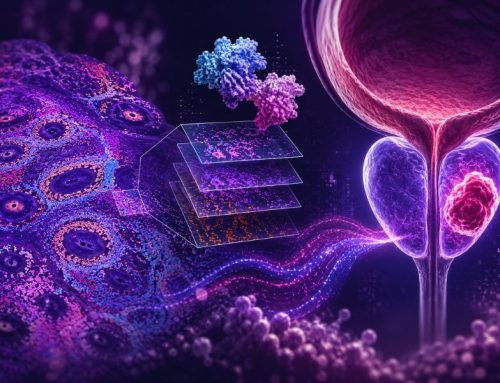
A recent single-cell analysis by researchers at KU Leuven and the VIB Center for Cancer Biology, spanning nine cancer types, has identified two distinct immune-reactive structures within tumors that predict both early and long-term responses to immune checkpoint blockade therapy. The researchers characterized over 600,000 cells to create a detailed atlas of the tumor microenvironment, revealing 70 shared cell subtypes and their spatial organization into functional “hubs” that could influence response to immunotherapy.
The study was published in Cell Reports Medicine.
Study Rationale
Predicting which patients will respond to immunotherapy remains a clinical challenge. Francesca Lodi, the first author of the study, explained that although previous pan-cancer single-cell studies have focused on individual cell types, this approach fails to capture how different immune populations interact within the tumor microenvironment. This limitation has hindered the development of biomarkers that reflect the complex cellular interactions driving immunotherapy response and resistance.
The research team aimed to address this gap by simultaneously profiling five cell types across 230 treatment-naive samples from 160 patients diagnosed with breast cancer, cervical carcinoma, colorectal cancer, glioblastoma, head and neck squamous cell carcinoma, hepatocellular carcinoma, ovarian carcinoma, melanoma, and non-small cell lung cancer.
Methodology
A methodological challenge was ensuring that cell-type proportions could be reliably compared across diverse tumor contexts.
“Some dissociation protocols may select certain cell types, while other protocols may select other cell types,” Lodi explained in an interview with Pathology News. “Using standardized dissociation protocols was therefore an important aspect of our study, as otherwise it might not have been possible to compare different cancer types.”
To validate their approach, the team directly compared cell-type fractions obtained via single-cell RNA sequencing against those estimated by deconvoluting paired bulk RNA-seq data from 25 samples across four cancer types. Although single-cell sequencing modestly enriched for immune cells, this enrichment was consistent across all cancer types, confirming that comparative analyses would be valid.
After applying Harmony for batch-effect correction and confirming its success using LISI scores, the researchers identified up to 70 cell subtypes shared across cancer types, including previously uncharacterized exhausted T cell populations.
Identification of Novel T Cell Subtypes
The researchers identified six subtypes of exhausted CD8+ T cells (TEX), including two previously unknown populations (CCL4+ and PKM+ TEX cells).
"Traditionally, TEX cells were often viewed as a single, uniform population," Lodi noted. "Our pan-cancer atlas challenged this view."
The CCL4+ TEX cells exhibited the highest expression of activation, TCR signaling, and exhaustion signatures, including the highest levels of PD-1 expression.
"This suggests that the abundance of CCL4+ TEX cells could serve as a highly specific biomarker for identifying tumors that are poised to respond to anti-PD1 therapy," Lodi explained.
In contrast, PKM+ TEX cells showed high expression of glycolysis-related genes alongside immune checkpoints LAG3 and TIGIT. These findings suggest that some patients might benefit from combination strategies where checkpoint blockade is paired with agents that modulate T cell metabolism.
The team also identified six CD4+ regulatory T cell (TREG) subtypes, ranging from resting to activated states, with TNFRSF9+ TREG cells showing increased exhaustion markers consistent with their heightened activation status.
Two Immune-Reactive Hubs
The team conducted pairwise correlation analyses of cell subtype abundances and identified two groups of strongly co-occurring subtypes, which they termed 'hubs.' The first hub resembled tertiary lymphoid structures (TLS), consisting of differentiated B cells (including four plasma cell subtypes, plasmablasts, and regulatory B cells), interferon-responsive macrophages, quiescent dendritic cells, and CD4+ T follicular helper cells.
"The TLS hub acts like the tumor's own immune campus," Lodi stated. "Spatially, it's organized into discrete, tightly clustered structures, like tiny lymph nodes forming right inside the tumor. Functionally, it's focused on adaptive immune education and maturation."
The cells within this hub rely heavily on chemokine signaling, particularly CXCL13, to organize and maintain their architecture.
The second hub represented a type-1 immunity response, comprising early inflammatory macrophages, immune-regulatory cells, antigen-presenting dendritic cells, and PD-1-expressing T cells, including CD8+ TEX cells and CD4+ T helper-1 cells.
"Spatially, it's more diffusely distributed throughout the tumor tissue, although it's often found next to the TLS hub," Lodi explained. "Functionally, this hub is built for immediate, direct anti-tumor execution."
The study showed extensive communication between subtypes within each hub, with 39 significant signaling pathways identified in the TLS hub and 72 in the type 1 immunity hub. The TLS hub showed prominent chemokine signaling, which is essential for immune cell migration, whereas the type 1 immunity hub was characterized by TNF and interferon signaling.
Spatial Validation Confirms Hub Architecture
To verify that these correlation patterns reflected actual spatial relationships, the team analyzed publicly available spatial transcriptomics data from 60 samples across six cancer types. The analysis confirmed that subtypes within each hub were spatially co-localized, although areas enriched for one hub did not necessarily overlap with those enriched for the other hub.
Multiplex immunohistochemistry on head and neck cancer sections provided additional validation at single-cell resolution, identifying specific cell types within each hub, including CD4+ T follicular helper cells, plasma cells, germinal center B cells, and regulatory B cells in TLS-enriched regions.
Hubs Predict Treatment Response
The team used a T cell reactivity score incorporating inhibitory markers (PD-1, CTLA-4, LAG3), cytotoxic markers (GZMB, PRF1), and activation markers, and found that subtypes within both hubs correlated positively with T cell reactivity across cancer types.
Long-term outcomes were evaluated using bulk RNA-seq data from 937 patients treated with immunotherapy in four clinical trials. Both hubs correlated with prolonged progression-free survival, with inflammatory macrophages, regulatory dendritic cells, and IgG-producing plasma cells being the primary drivers of these associations.
"You need the type-1 hub for the initial fight, and the TLS hub to keep the immune system educated and ready for the long haul," Lodi explained.
The type-1 immunity hub signals tumors with activated effector populations held back by checkpoints, whereas the TLS hub contributes to sustained, durable responses through continuous antigen recognition and presentation.
Moving Toward Neighborhood Biomarkers
The team developed an interactive application called Shiny that allows researchers and clinicians to explore the atlas, rank cancer types by specific gene signature expression, and correlate cell subtype abundances with custom signatures.
"This correlative power moves beyond simple single-gene biomarkers to focus on the entire TME niche, the distinct cellular ecosystems that drive or resist therapeutic response," Lodi said.
Users can identify "neighborhood biomarkers," such as the combined abundance of inflammatory macrophages plus exhausted T cells, rather than relying on single cell types.
The application enables hypothesis generation for combination therapies. If a tumor shows suppressed-but-ready immune infiltrates alongside specific oncogenic pathway activation, combining pathway inhibitors with checkpoint blockade becomes a testable clinical trial hypothesis.
Limitations and Future Directions
The authors acknowledge that the sample sizes for some cancer types were relatively small, that the Visium platform lacks single-cell resolution, and multiplex immunohistochemistry was limited to 60 antibodies.
"The next step of a future atlas involves generating a spatial tumor map at true single-cell resolution to integrate the deep cell refinement we achieved based on scRNA-seq with accurate spatial locations," Lodi noted.
Technologies like Xenium (10x Genomics) and MERSCOPE (Vizgen) are being deployed to achieve this level of detail.
"The key to this study was the coordination among clinicians, lab technicians, and bioinformaticians required to generate such a large, homogeneous dataset entirely in-house," Lodi concluded. "This unique effort has yielded an invaluable resource for the entire scientific community, providing a strong foundation for future studies aiming to understand TME heterogeneity and its implications in therapy response and resistance."
The study received financial support from VIB Grand Challenges.
References
- Lodi F, Vanmassenhove S, Chen D, et al. Decoding tumor heterogeneity: A spatially informed pan-cancer analysis of the tumor microenvironment. Cell Rep Med. 2025;6(10):102416. doi:10.1016/j.xcrm.2025.102416
No audio available for this article yet.
No quiz available for this article yet.