Author: Sidney Ocanagil-Tunstall
Perihilar cholangiocarcinoma (PHCC), also known as Klatskin tumors, represents one of the most challenging malignancies in oncology. With an incidence rate of 1-2 cases per 100,000 individuals in the Western world, this rare cancer is characterized by late diagnosis and poor long-term survival rates, even with curative surgical treatment. Artificial intelligence (AI) has emerged as a promising tool in oncology, particularly through AI-based digital histopathology, offering the potential to improve prognostic predictions in various cancers, including PHCC. However, while AI offers significant advancements, its role in PHCC remains a step forward, but not yet the leap required for clinical impact.
Background on Perihilar Cholangiocarcinoma
Perihilar cholangiocarcinoma arises at the junction of the right and left hepatic bile ducts. Surgical resection, often combined with complex liver and vascular procedures, is currently the most effective curative treatment. However, even among patients who undergo surgery, survival rates remain dismal, ranging from 13.5% to 42% at five years. Several clinical and pathological factors have been identified as prognostic indicators, but predicting individual patient outcomes remains difficult.
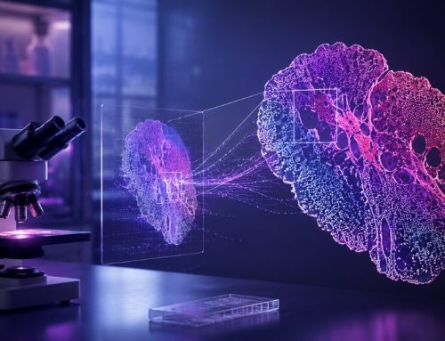
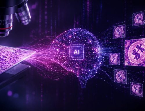
Given the highly heterogeneous nature of PHCC and its morphological complexity, standard clinical assessments fall short of providing precise survival predictions. This gap in prognosis has motivated researchers to explore AI-based digital histopathology as a novel solution. AI, specifically convolutional neural networks (CNNs), can analyze whole-slide images (WSI) from pathology samples, identifying patterns and features beyond what is visible to the human eye. These models promise to augment traditional methods, providing pathologists with insights that may improve individual patient management.
Study Objectives and Methodology
In this study, researchers from the University Hospital of Essen sought to determine whether AI-based histopathological analysis, combined with clinical data, could improve survival predictions in PHCC patients.¹ A cohort of 142 patients who underwent surgical treatment for PHCC was retrospectively analyzed. Clinical data were collected, including tumor size (T), grade (G), and intraoperative factors, such as transfusion requirements.
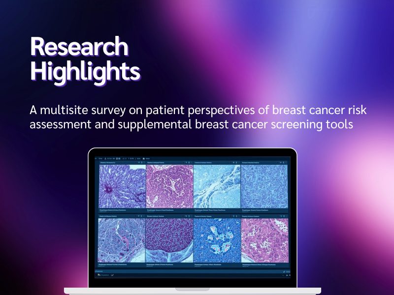
The research team utilized CNN-based models to analyze WSIs, identifying regions of interest, such as tumor nests and malignant cells, to extract features. These CNN features were then integrated with clinical factors in survival models, with the goal of improving prognosis prediction.
Results and Key Findings
The study revealed that, while the CNN-based models successfully identified certain histopathological features, their integration with clinical data did not significantly improve survival predictions compared to clinical data alone. The most reliable independent predictors of survival remained tumor grade, tumor size, and intraoperative transfusion requirements. Despite the promise of AI in histopathology, the AI-based models failed to substantially enhance prognosis accuracy.
One of the more successful aspects of the CNN models was their ability to generate heatmaps that highlighted areas of interest for pathologists. These heatmaps acted as visual aids, assisting pathologists in identifying potential tumor regions. However, these heatmaps were not precise enough to replace the expertise of an experienced hepatobiliary pathologist and were, at best, a supportive tool for less experienced clinicians.
Challenges and Limitations of AI in PHCC
Several factors contributed to the limited success of the AI-based models in this study. First, the complex and variable morphology of PHCC tumors presented challenges in feature extraction. While the CNN models could identify general areas of tumor cells, they lacked the precision required to capture the nuanced histopathological characteristics needed for prognosis prediction.
Moreover, the study’s methodology involved annotating tumor areas rather than individual tumor cells, leading to potential inaccuracies in the extracted features. This approach likely resulted in the inclusion of peritumoral tissue in the analysis, which may have diluted the model’s ability to distinguish relevant prognostic features.
The black-box nature of CNNs also posed interpretability challenges. While the models could predict outcomes based on the extracted features, understanding the exact rationale behind their decisions remains difficult, limiting their clinical utility.
The Path Forward: Improving AI Models for PHCC
Despite these limitations, the study provides a proof of concept for AI’s potential in digital histopathology. The results suggest that while current AI-based models are not yet clinically relevant, they offer a foundation for future improvements. Enhancing the accuracy of tumor cell identification, expanding the dataset to include more diverse cases, and refining feature extraction techniques are crucial next steps.
Future research should explore multicentric collaborations to increase the volume of data available for training AI models. Additionally, adopting more sophisticated techniques for data augmentation, normalization, and pretraining could enhance the models’ generalizability and performance.
Conclusion
AI-based digital pathology is a promising tool in the fight against PHCC, but the current models represent a step, not a leap, towards improving prognosis predictions. While CNN-generated heatmaps can aid in histopathological assessments, further refinement is needed before AI can be relied upon for accurate clinical decision-making in PHCC. For now, AI remains a complementary tool, one that may assist pathologists in identifying areas of interest, but it is not yet capable of replacing human expertise in this highly complex malignancy.
Future Directions
The journey to fully integrating AI into the clinical management of PHCC is ongoing. Researchers and clinicians alike must continue to refine AI models, ensuring they become robust, interpretable, and clinically valuable tools. A larger, multicentric dataset and a shift towards more precise tumor cell annotations could mark the beginning of this next step forward, as the oncology community works to improve outcomes for patients with this devastating disease.
References
This article is based on and incorporates elements from the following open access research paper, licensed under the Creative Commons Attribution 4.0 International License. The original paper has been remixed, transformed, and built upon for this work.
- Hoyer DP, Ting S, Rogacka N, Koitka S, Hosch R, Flaschel N, Haubold J, Malamutmann E, Stüben BO, Treckmann J, Nensa F, Baldini G. “AI-based digital histopathology for perihilar cholangiocarcinoma: A step, not a jump.” Journal of Pathology Informatics. 2024;15:100345. ISSN 2153-3539. https://doi.org/10.1016/j.jpi.2023.100345. Link to article.
No audio available for this article yet.
No quiz available for this article yet.