DP65
by Christos Evangelou, MSc, PhD – Medical Writer and Editor
In a new study, researchers at NYU Grossman School of Medicine and Warren Alpert Medical School of Brown University investigated the relationship between digital pathology features from whole slide images (WSIs) of core needle biopsies and radiomic features from post-neoadjuvant chemotherapy magnetic resonance imaging (MRI) scans in patients with triple-negative breast cancer (TNBC).1
The study uncovered novel correlations between histological features from digital pathology and radiomic texture patterns on MRI that are associated with treatment response in patients with TNBC. These findings suggest that the integration of digital pathology and MRI may provide a better understanding of the tumor microenvironment in TNBC and inform treatment strategies for this aggressive breast cancer subtype.
“Our findings highlight the potential of using digital pathology and radiomics to predict which TNBC patients are likely to achieve pathologic complete response after neoadjuvant chemotherapy,”
said Sean M. Hacking, MD, who is a clinical assistant professor at NYU Grossman School of Medicine and the first author of the study.
“This could lead to more personalized treatment plans, optimizing the effectiveness of neoadjuvant chemotherapy and potentially sparing patients from unnecessary treatment side effects.”
The report was published in Breast Cancer.
Unmet Need: Decoding the Tumor Microenvironment in TNBC
TNBC is an aggressive and difficult-to-treat form of breast cancer characterized by the absence of three receptors that serve as therapeutic targets. Therefore, patients with TNBC have limited treatment options and poor outcomes.
Despite recent advances in characterizing the tumor microenvironment in various cancer types, tumors in patients with TNBC are highly heterogeneous. Our understanding of the complex interplay between cancer cells and their surrounding microenvironment in patients with TNBC remains limited. This knowledge gap has
hindered the development of targeted therapies for this aggressive subtype of breast cancer.
“Our motivation for conducting this study stemmed from the need to improve our understanding of TNBC and to identify new ways to predict how patients will respond to neoadjuvant chemotherapy,”
noted Dr. Hacking.
Bridging the Gap: A Multidisciplinary Approach
To address this knowledge gap, a multidisciplinary team of researchers integrated two advanced technologies: digital pathology and radiomics. By analyzing WSIs from core needle biopsies and MRI scans of 12 patients with TNBC, the team sought insights into the intricate relationships between the tumor’s histological features and its radiological characteristics.1
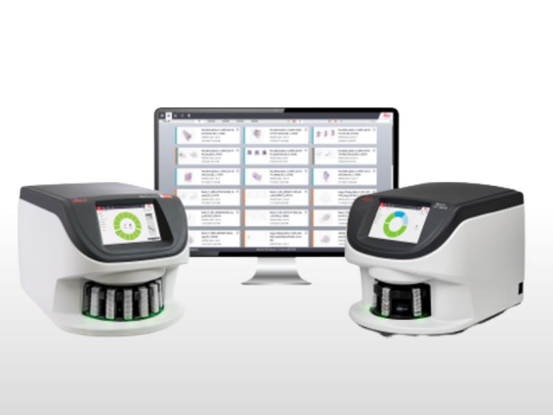
Tissue slides from patients with TNBC were digitized using a high-resolution scanner, enabling detailed quantitative analysis through machine learning algorithms. These algorithms were trained to identify and quantify various components of the tumor microenvironment, including tumor cells, collagen, myxoid stroma, and immune cells.
Moreover, the team annotated post-neoadjuvant chemotherapy MRI scans to extract radiomic features using advanced computational pipelines. These features quantified aspects such as tumor shape, texture, and intensity, providing a comprehensive characterization of the radiological profile of the tumor.
The team also analyzed the relationship between digital pathology features from WSI and radiomic features from MRI scans, focusing on how these features correlate with pathologic complete response (pCR) after neoadjuvant chemotherapy.
“By exploring the correlation between digital pathology, radiomics, and pCR, we aimed to uncover insights that could lead to better patient outcomes,”
explained Dr. Hacking.
Significant Associations Between Histological Characteristics and Radiomic Features of TNBC Tumors
The researchers identified several significant associations between histological characteristics on WSI and the radiomic features of TNBC tumors. For instance, high proportions of collagenous stroma in the tumor microenvironment were associated with high homogeneity in the appearance of tumors on MRI, as measured by the size zone non-uniformity normalized (SZNN) feature.1 Conversely, high tumor cell proportions correlated with low tumor homogeneity, suggesting a more chaotic and disorganized tumor structure.
The study also demonstrated that high levels of myxoid stroma on WSI were associated with low similarity in gray-level intensity values on MRI, as captured by the gray-level non-uniformity (GLN) feature.1 Furthermore, increased immune cell infiltration was associated with a high proportion of smaller, darker zones within the tumor, as quantified by the small area low gray-level emphasis (SALGE) feature.
Potential Implications: Tailoring Treatments and Improving Outcomes
The correlations between digital pathology and radiomic features reported in this study have potential implications for the management of TNBC. By gaining a deeper understanding of the tumor microenvironment and its radiological features, clinicians could better tailor treatment strategies to individual patients.
For instance, the study identified specific histological characteristics on WSIs and radiomic features associated with pCR, a crucial indicator of treatment success in patients with TNBC. Lower collagen and myxoid stroma levels and high tumor cell proportions on WSI were associated with response (i.e., pCR) to neoadjuvant chemotherapy. Low GLN and high SZNN on MRI were also correlated with pCR after neoadjuvant chemotherapy.1
“These correlations suggest that integrating digital pathology and radiomics could enhance our ability to predict treatment outcomes in TNBC,”
noted Dr. Hacking.
He explained that, by integrating histologic characteristics and radiomic features, oncologists could potentially adjust neoadjuvant chemotherapy regimens or explore complementary targeted therapies that target specific components of the tumor microenvironment, such as the stromal compartment or immune cells. This personalized approach could improve treatment efficacy, minimize side effects, and ultimately enhance patient outcomes.
According to Dr Hacking, the most innovative aspect of this work is
“the demonstration of a clear, quantifiable relationship between digital pathology and radiomic features in predicting pCR in TNBC.”
Looking Ahead
“While our study provides important insights, questions remain regarding the broader applicability of our findings across different populations and cancer subtypes,”
Dr. Hacking acknowledged.
Limitations of this study include the small sample size and the use of a single biopsy, which may not fully capture the heterogeneity within the tumor. Larger validation studies are needed.
“Future studies will need to involve larger patient cohorts and explore the integration of these predictive models into clinical practice. Additionally, further research is needed to understand the underlying biological mechanisms driving the correlations we observed,”
said Dr. Hacking.
Additionally, exploring the potential integration of other advanced imaging modalities, such as positron emission tomography, could further enrich the understanding of the tumor microenvironment and its radiological signatures.
Despite these limitations, Dr Hacking believes that this new multidisciplinary approach represents a significant step forward in the precision medicine landscape for TNBC.
“We believe our study opens up new avenues for improving the precision of TNBC treatment strategies and are excited about the potential impacts of our findings on patient care,”
he said.References
1. Hacking SM, Windsor G, Cooper R, Jiao Z, Lourenco A, Wang Y. A novel approach correlating pathologic complete response with digital pathology and radiomics in triple-negative breast cancer. Breast Cancer. Published online February 13, 2024. doi:10.1007/s12282-024-01544-y
No audio available for this article yet.
No quiz available for this article yet.