by Christos Evangelou, MSc, PhD – Medical Writer and Editor
A recent study from Paige (New York, NY, US) and New England Pathology Associates (Springfield, MA, US) demonstrates that artificial intelligence (AI) can significantly enhance the ability of pathologists to detect breast cancer metastases in lymph nodes. Developed by Paige.AI, the AI tool not only improved diagnostic accuracy but also drastically reduced the time required for analysis.1
According to the authors, this advancement could lead to more efficient and reliable breast cancer staging, potentially improving treatment decisions and patient outcomes.
The report was published in the American Journal of Surgical Pathology.
Challenges in Lymph Node Assessment
Breast cancer staging relies heavily on accurate detection of lymph node metastases. However, lymph node assessment is challenging and time consuming.1 Previous studies have shown that up to 24% of patients may have their nodal stage changed when lymph nodes are re-examined by specialized breast pathologists.2 According to the authors, this discrepancy highlights the need for tools to improve the accuracy and efficiency of lymph node assessment.
“We know that pathologists are not perfect at breast cancer lymph node assessment,” said Dr. Juan Antonio Retamero, Medical Vice President and Medical Director at Paige. “The literature shows that a significant proportion of patients examined by general pathologists have their nodal stage changed, usually upstaged, when the cases are re-reviewed by breast specialist pathologists.”
Dr. Retamero and colleagues set out to address this challenge by investigating how an AI-assisted approach could impact the performance of pathologists in detecting breast cancer metastases in lymph nodes.
Methodology
The researchers evaluated the impact of AI assistance on the diagnostic performance of pathologists. To this end, they used Paige BLN, an AI algorithm trained on over 32,000 breast sentinel lymph node whole slide images (WSIs) from more than 8,000 patients.1 This tool highlights areas suspicious for metastasis on digital slides.
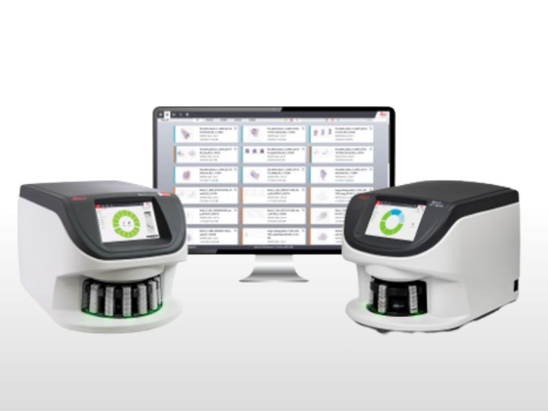
Commenting on the integration of the AI system into the existing workflow of pathologists, Dr. Retamero stated that Paige BLN is integrated into the FullFocus viewer, an FDA-approved digital pathology viewer that offers all the functionalities that pathologists need for their day-to-day diagnostic needs.
“Paige BLN is easy and intuitive to use, and full training is provided. This lasts approximately one hour, after which pathologists are ready for its use,” he added.
The team assembled a dataset of 167 WSIs of breast sentinel lymph nodes from 148 patients. This included 98 benign slides and 69 slides with metastases of varying sizes, including isolated tumor cells (ITCs), micrometastases, and macrometastases.1
Three board-certified pathologists with 21–32 years of experience participated in the study. They reviewed the dataset twice: once without AI assistance and once with AI assistance (after a 3-week washout period).1 The researchers recorded the diagnoses and the time taken to review each slide. They then compared the accuracy and efficiency of diagnosis between the AI-assisted and unassisted readings.
AI Assistance Improves Diagnostic Accuracy
The study demonstrated significant improvements in both diagnostic accuracy and efficiency when pathologists used the AI tool. Overall sensitivity increased from 81.2% (95% confidence interval [CI], 67.6%–94.7%) without AI to 93.2% (95% CI, 87.7%–98.8%) with AI assistance.1 Two of the three pathologists showed statistically significant improvements in sensitivity, rising from the 70% range to over 90%.
Dr. Retamero explained that improvements in metastasis detection were greatest for pathologists who took the shortest time to evaluate the dataset, which suggests that pathologists who took the longest time were the most accurate in their diagnosis.
“But in today’s busy pathology practices, pathologists rarely have the luxury of time,” he added.
The most substantial improvements were seen in detecting ITCs and small micrometastases, which, according to the authors, are typically the most challenging to identify.1
“We saw a significant improvement in the detection of metastases, and this was greatest for ITCs; however, these have relatively little clinical relevance, except in cases treated with neoadjuvant therapy,” noted Dr. Retamero.
He added, however, that the observed improvements in the detection of micrometastases are clinically significant, as they can affect the nodal stage of the patient.
The Paige BLN algorithm demonstrated a standalone sensitivity of 92.8% and specificity of 94.9%.1 Notably, the AI system achieved a 78% sensitivity for detecting ITCs, which, according to the authors, is superior to rates typically reported in the literature.
AI Assistance Improves Diagnostic Efficiency
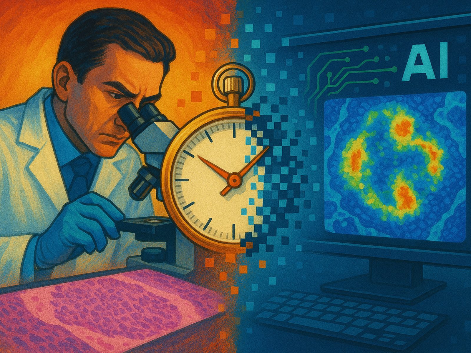
The average time to review a slide decreased from 128.5 seconds without AI to 58.3 seconds with AI assistance, which represents a 55% reduction (P < 0.001). Time savings were observed for both benign and malignant slides.1 The most significant time reduction (75.4%) was seen for large micrometastases, decreasing from 79.2 seconds to 19.5 seconds per slide.
“Pathologists became not only more precise in their diagnosis but also twice as fast when they were aided by AI,” emphasized Dr. Retamero. “Even those pathologists who were very precise, but at the cost of time, saw their performance significantly speeded up.”
Future Work
The authors acknowledge that with only three participating pathologists, larger studies are needed to confirm the generalizability of the findings.1 Moreover, the majority of cases were treatment-naive ductal carcinomas, and further research is needed to evaluate the performance of the AI system on a broader range of breast cancer types and post-treatment cases.1
Commenting on the potential future applications of AI in pathology beyond lymph node metastasis detection, Dr. Retamero said:
“We are seeing the advent of foundation models in AI that are capable of multiple downstream tasks, ranging from tumor detection, of pretty much any tumor type from any origin, from any cell lineage to digital biomarker or clinical outcome prediction.”
This study was funded by Paige, and no specific grant was received from any funding agency in the public or not-for-profit sectors.
References
- Retamero JA, Gulturk E, Bozkurt A, et al. Artificial Intelligence Helps Pathologists Increase Diagnostic Accuracy and Efficiency in the Detection of Breast Cancer Lymph Node Metastases. Am J Surg Pathol. 2024;48(7):846-854. doi:10.1097/PAS.0000000000002248
- Vestjens JHMJ, Pepels MJ, de Boer M, et al. Relevant impact of central pathology review on nodal classification in individual breast cancer patients. Ann Oncol. 2012;23(10):2561-2566. doi:10.1093/annonc/mds072
No audio available for this article yet.
No quiz available for this article yet.