AIRAQc – Automated Quality Control for WSI
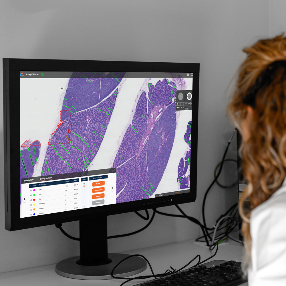
AIRAQc automates digital pathology preanalytical quality checks by using AI to identify and quantify artifacts. It automatically accepts or flags images for reprocessing or rescanning based on predefined thresholds set by the user, eliminating the need for manual analysis. This streamlines workflows, saves resources, and ensures consistent image and data quality for accurate analysis in your pathology laboratory.

Identification and Quantification of Artifacts in WSIs
Ensure Only High-Quality WSIs Reach Your IMS
Benefits of AIRAQc:
Enables 100% QC across data
- Locate, annotate and quantify areas in the data associated with artifact and missing tissue
Standardization across the QC process
- Removes intra and interobserver variability associated with manual QC
- Around-the-clock QC with email notifications, and remote access
Dashboard Data
- Aggregate and communicate results across the enterprise
- Individual scanner QC data
- Annotations and associated data
Reduces downstream problems associated with artifacts and missing tissue
- Pathologists time
- Misdiagnosis
- Errors associated with feeding artifacts to AI and image analysis applications
- Blur is difficult and time-consuming to locate at different zoom levels
Frees time for Histotechnicians
Key Capabilities
Advanced Artifact Detection
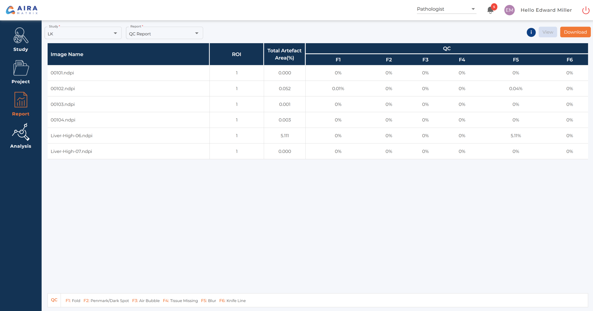
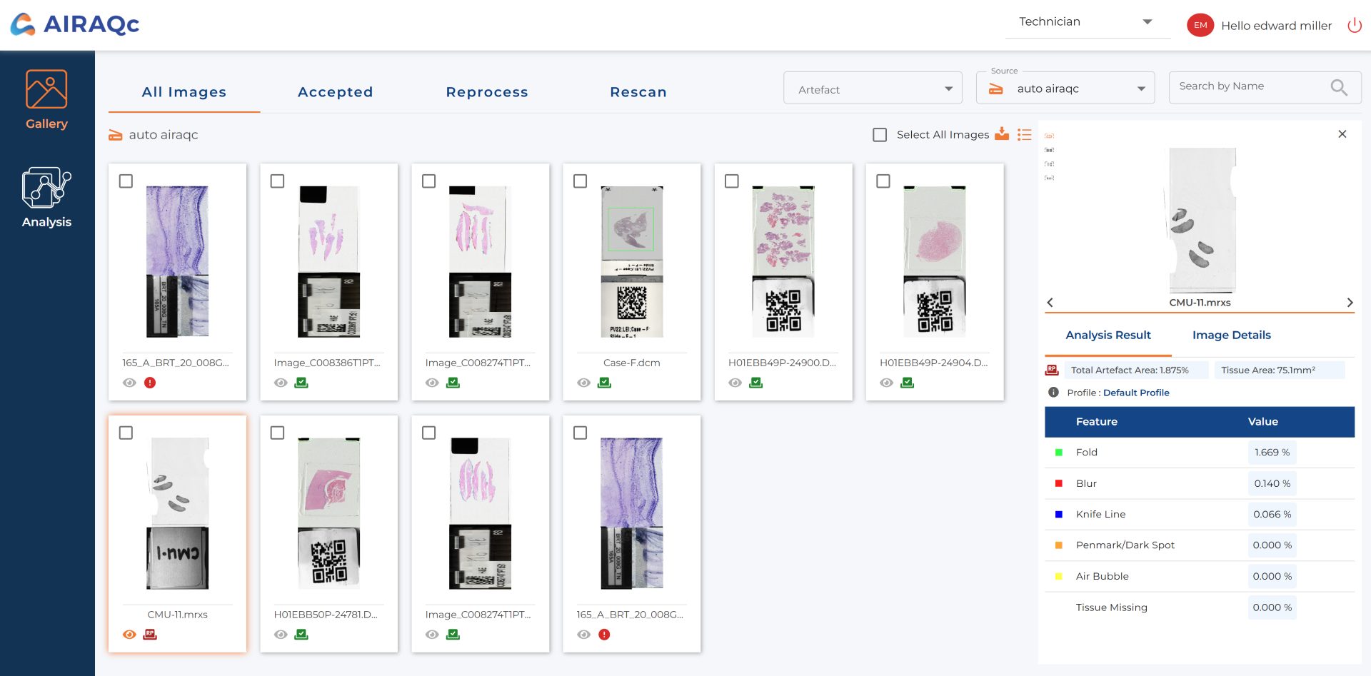
Processing Artifacts: Tissue folds, pen marks, air bubbles, out of focus, knife lines, dark spots, missing tissue
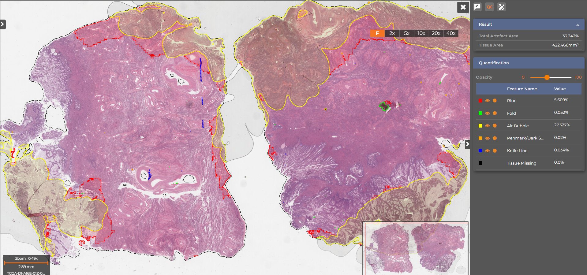
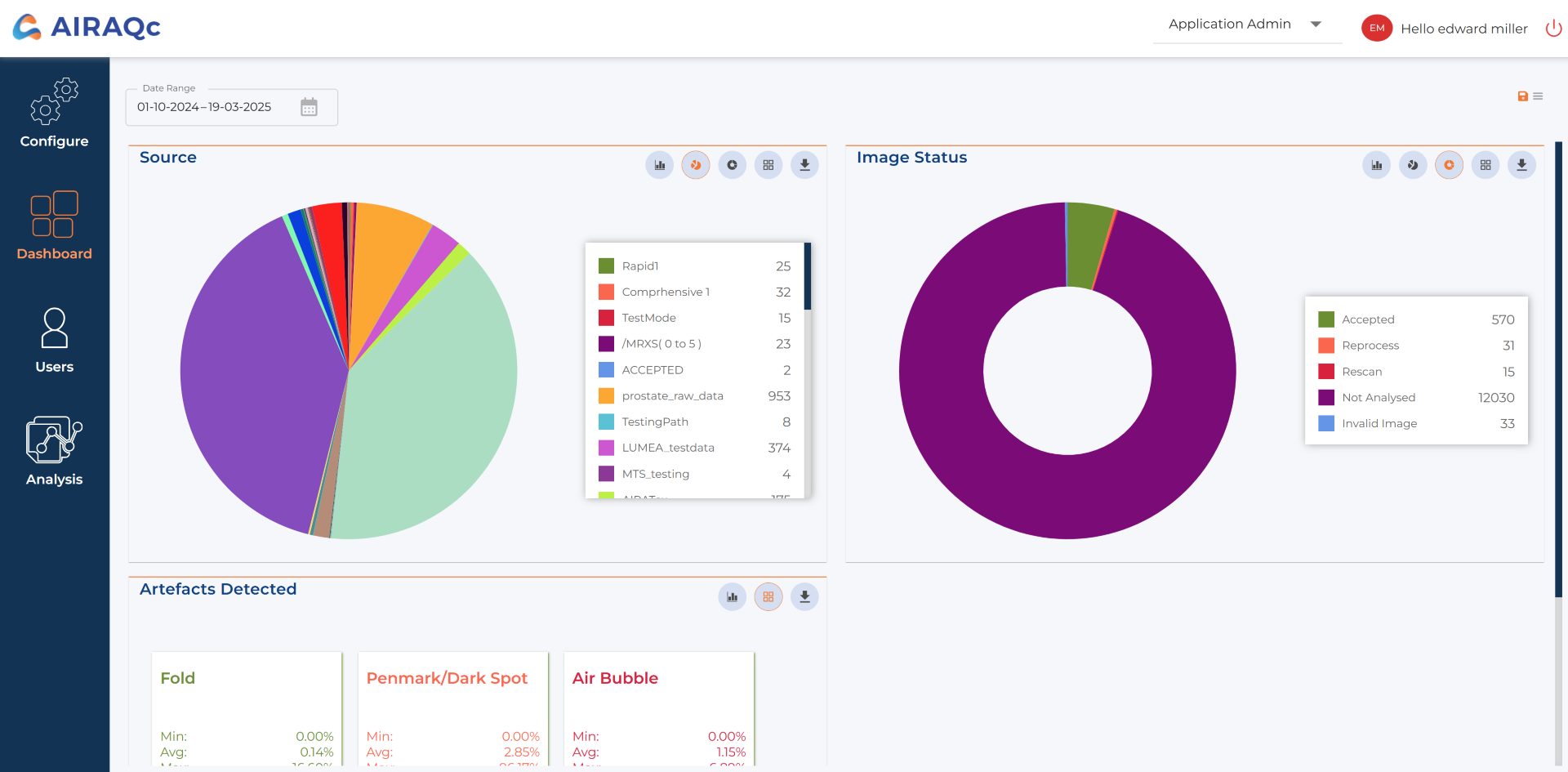
Scanning Artifacts: Out-of-focus areas, missing tissue, no tissue detected

Automated QC with Customizable Thresholds

Flag images as Accept, Reprocess, or Rescan based on user-defined thresholds
Process 10 WSIs per minute (1GB each) with 95% sensitivity and 96% specificity
Real-Time Analytics & Compliance Support
Detailed artifact reports for root cause analysis & corrective actions
A centralized dashboard for lab-wide QC insights
Supports CLIA compliance with full audit traceability

Seamless Integration & Flexible Deployment

Works with LIS, IMS, and PACS, ensuring no workflow disruption
Compatible with all major WSI formats
On-premise or Cloud Deployment, tailored to pathology lab setup
| Supported Stains | H&E, IHC |
|---|---|
| Species | Agnostic |
| Artifacts Detected | Tissue folds, pen marks/ dark spots, air bubbles, coverslip lines, knife lines, out of focus, missing tissue, no tissue |
| Analysis Mode | Auto, Manual |
| Throughput | 10 WSIs per minute (1GB each) |
| Deployment | Cloud, On-Premise |
| Output Format | CSV, Excel |
| Scanner Compatibility | Compatible with multiple image formats and scanners |
More Solutions from AIRA Matrix
Compare Solutions
Get Pathology News Delivered to Your Inbox
Learn More
AIRA Matrix News
Mastering Image Quality Control with AIRA Matrix
Editor2026-05-22T14:29:30+00:00November 18, 2025|
AIRA Matrix Partners with Gestalt to Elevate Diagnostics with AI-Driven QC Platform
admin@pathologynews.com2025-06-06T10:15:48+00:00March 28, 2025|
AIRA Matrix and PathNet Have Joined Forces to Bring to Life a Highly Progressive Digital Platform for Prostate Cancer
admin@pathologynews.com2025-06-06T10:16:10+00:00March 27, 2025|
Press Release: AIRA Matrix, Lumea, and Acupath Have Partnered to Provide a Diagnostic AI Solution for the URO17® Bladder Cancer Biomarker
admin@pathologynews.com2025-06-06T10:16:47+00:00March 26, 2025|


Reviews
There are no reviews yet.