by Christos Evangelou, MSc, PhD – Medical Writer and Editor
In a recent study, researchers at the University of Chicago and the University of Chicago Medical Center developed a low-cost, open-source digital pathology workstation that performs on par with expensive clinical-grade systems in cancer diagnostics.1 This $230 device, coupled with artificial intelligence, could increase access to advanced cancer care in resource-limited settings, potentially addressing the growing cancer burden in low- and middle-income countries.
The report was published in eBioMedicine.
Bridging the Diagnostic Gap
The increasing incidence of cancer in low- and middle-income countries raises the need for accessible and accurate diagnostic tools. Although cancer cases in low Human Development Index (HDI) countries are projected to double between 2008 and 2030, these regions often lack access to the advanced diagnostic technologies that have become standard in high-income nations.1
Dr. Alexander T. Pearson, the study’s corresponding author, explained the motivation behind their work:
“With the global burden of cancer shifting towards low- and middle-income countries, adapting the diagnostics tools for use in a wider range of clinical settings will be essential for keeping up with a coming wave of cancer diagnoses around the world.”
Digital pathology has emerged as a powerful tool in cancer diagnostics. However, the high cost of equipment has put this technology out of reach for many healthcare providers in resource-limited settings.1 The researchers set out to address this disparity by developing an open-source, low-cost alternative that could match the performance of high-end systems.
“The personnel and equipment shortages that our colleagues and collaborators face in different parts of the world create situations in which automation of cancer diagnostics can be particularly useful,” Dr. Pearson noted. “Because of this, we wanted to build an accessible workstation that could serve as an end-to-end platform for deep learning classification of tissue samples.”
Building an Affordable Digital Pathology System
The team modified the open-source OpenFlexure Microscope v6 design, creating a 3D-printed device that costs just $230. This device was paired with a low-cost Raspberry Pi computer and camera module.1
The researchers developed Slideflow, an open-source software package for deep learning analysis of digital pathology images. This software can run on the low-power Raspberry Pi, eliminating the need for expensive computational hardware.1
The team also trained artificial intelligence models to classify different types of cancer, including head and neck squamous cell carcinoma (HNSCC), lung cancer, and breast cancer subtypes.
Testing the System
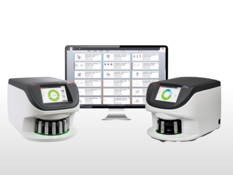
To evaluate their low-cost system, the researchers compared its performance against a clinical-grade Aperio AT2 digital pathology microscope, which costs around $250,000.1 They tested the system’s ability to predict HPV status in HNSCC, distinguish between lung adenocarcinoma and squamous cell carcinoma, and differentiate between invasive ductal and invasive lobular breast carcinoma.1
The team used publicly available data from The Cancer Genome Atlas (TCGA) to train the deep learning models and validated them on a small set of samples from the University of Chicago Medical Center.1
Low-Cost Digital Pathology Performs On Par With High-Cost System
Despite the significant cost difference, the low-cost system performed comparably to the high-end Aperio AT2 in all three classification tasks. When used to predict HPV status in HNSCC, the low-cost system achieved a patient-level area under the receiver operating characteristic curve (AUROC) of 0.88, compared to 0.84 for the Aperio AT2.1
The low-cost system also achieved high patient-level classification in distinguishing between adenocarcinoma and squamous cell carcinoma, with an AUROC of 1.0. The patient-level AUROC for the model tested on images captured with the high-cost Aperio AT2 system was 0.7.1
In addition, the low-cost system achieved a patient-level AUROC of 0.80, compared to 0.84 for the Aperio AT2, in differentiating between invasive ductal and invasive lobular carcinoma.1 Importantly, there were no statistically significant differences in performance between the two systems for any of the cancer subtypes tested.
According to the authors, the ability to maintain model performance despite decreased image quality and low-power computational hardware demonstrates that it is feasible to reduce costs associated with deploying deep learning models for digital pathology applications.
Dr. Pearson commented on the implementation of their technology:
“Because the workstation we developed is open-source and modular, we envision it being modified and developed according to the needs of the clinicians using them. For example, if a lab already has access to imaging technology but does not have enough pathologists, any image can theoretically be uploaded into our deep learning algorithm for classification. On the other hand, a low-cost image of a patient’s tissue sample could be captured using our 3D-printed microscope and sent to a remote collaborator for analysis.”
Limitations and Future Directions
Although the results are promising, the researchers acknowledge several limitations of their study. The external validation was performed on a limited number of samples (10 per cancer type), and larger studies are needed to confirm the system’s performance.1 In addition, the study focused on only three specific cancer classifications. Further research is needed to validate the system for other cancer types and diagnostic tasks. Moreover, the training data primarily came from patients in the United States and Europe. Ensuring the system works well on diverse patient populations is crucial for global implementation.
Divya Choudhury, the first author of the study, commented on these limitations:
“We have not tested the ability to use the images captured by the low-cost device to carry out more difficult classification tasks, such as hormone receptor status or microsatellite instability. That said, having a low-cost automated platform that can detect something as straightforward as cancer vs non-cancer may be extremely useful clinically in places without access to robust pathology services.”
Choudhury outlined their next steps, which include further validation of the system with more classification tasks, multiclass models, and more representative datasets.
“As deep learning continues to improve and gain approval for clinical use, clinical trials that focus on cost-reduction to improve the accessibility of this technology will be essential,” she added.
Commenting on the role of open-source tools in the future of precision oncology, Choudhury noted:
“Open-source tools are very valuable for the equitable deployment of new health technologies. Keeping tools open source helps make them available to adapt and modify to best suit the needs of the end user, including for patients and providers with limited resources.”
Dr. Pearson concluded with his vision of how the implementation of this technology could improve access to diagnostic tools in limited-resource regions:
“We hope that this inspires other researchers to invest in efforts to reduce cost and improve accessibility so that the most cutting-edge technology can be available to as many communities around the world as possible, and not restricted to the most well-resourced settings as they primarily are today.”
This study received financial support from the National Institutes of Health, National Cancer Institute, National Institute of Dental and Craniofacial Research, Stand Up to Cancer Fanconi Anemia Research Fund– Farrah Fawcett Foundation Head and Neck Cancer Research Team Grant, and the European Union Horizon Program.
References
- Choudhury D, Dolezal JM, Dyer E, et al. Developing a low-cost, open-source, locally manufactured workstation and computational pipeline for automated histopathology evaluation using deep learning. EBioMedicine. 2024;107:105276. doi:10.1016/j.ebiom.2024.105276
No audio available for this article yet.
No quiz available for this article yet.