The following abstract is drawn from a recently published paper in the National Library of Medicine. We invite you to read the full paper and join the conversation, become a member of the Pathology News community to share your thoughts, ask questions, and engage with others around this work.
Authors: Shuwei Zhang#,1, Houpu Yang#,1, Yiyin Zhang#,1, Xiaoxian Li2, Jin Zhao1, Yuanyuan Zhang3, Ping Xue4, Hua Kang5, Hongchuan Jiang6, Wenhui Ren7, Shu Wang1
Abstract
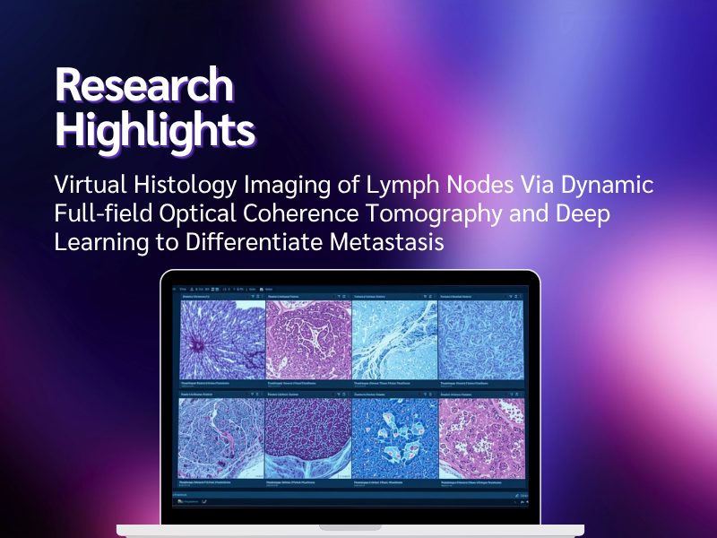
Objective: The current pathological diagnosis of lymph node metastasis is time-consuming, labor-intensive, and dependent on sectioning of paraffin blocks. Herein, in a prospective cohort of patients with breast cancer, we validated dynamic full-field optical coherence tomography (D-FFOCT), a virtual pathology tool integrating deep learning for nodal metastasis detection, and offering rapid and label-free histologic approximations of fresh tissues.
Methods: In a prospective dual-center cohort of 155 patients with breast cancer, 747 freshly bisected lymph node slides were obtained via D-FFOCT. Surgeons interpreted each slide with histopathology as the gold standard. A deep learning model was trained on 28,911 patches (corresponding to 590 slides) and tested on 7,736 patches (corresponding to 157 slides). The results were mapped to the slide level for potential intraoperative evaluation.
Results: D-FFOCT strongly correlated with hematoxylin and eosin (H&E)-stained histological images. Surgeons achieved 97.10% specificity in nodal diagnosis with D-FFOCT. The performance of the artificial intelligence (AI) model was not inferior to that of human experts and had a sensitivity/specificity of 87.88%/91.94% and an area under the receiver operating characteristic curve of 0.899 at the slide level. The human-AI collaborative system reduced labor requirements by 75% and increased the specificity by 6.5%, to 98.39%.
Conclusions: D-FFOCT has excellent potential as a tool for assessing lymph node metastatic status without tissue preparation or consumption. The integration of D-FFOCT with deep learning decreases labor demands and maintains high accuracy, thereby enabling streamlined nodal prediction independent of routine pathology procedures.
Read the full article: Virtual histology imaging of lymph nodes via dynamic full-field optical coherence tomography and deep learning to differentiate metastasis – PubMed
1Breast Center, Peking University People’s Hospital, Beijing 100044, China.
2Department of Pathology and Laboratory Medicine, Emory University, Atlanta, GA 30322, USA.
3Department of Pathology, Peking University People’s Hospital, Beijing 100044, China.
4Department of Physics and State Key Laboratory of Low-dimensional Quantum Physics, Tsinghua University, Beijing 100084, China.
5Department of General Surgery, Xuanwu Hospital, Capital Medical University, Beijing 100053, China.
6Department of Breast Surgery, Beijing Chaoyang Hospital, Capital Medical University, Beijing 100020, China.
7Department of Clinical Epidemiology and Biostatistics, Peking University People’s Hospital, Beijing 100044, China.
No audio available for this article yet.
No quiz available for this article yet.