by Christos Evangelou, MSc, PhD – Medical Writer and Editor
A new automated imaging platform developed at Georgia Institute of Technology and Emory University could make advanced spatial biology techniques accessible to laboratories that have limited resources. The imaging platform, called PRISMS (Python-based Robotic Imaging and Staining for Modular Spatial Omics), combines open-source software, affordable liquid handling robots, and standard microscopy equipment to enable researchers to visualize RNA and proteins within tissues at single-molecule resolution at a fraction of the cost of commercial alternatives.
The study was published in Lab on Chip.
Study Rationale
Spatial omics technologies remain out of reach for many laboratories and research institutes. Typically, commercial spatial omics platforms require the use of costly proprietary instrumentation, specialized reagents, and rigid workflows that offer little room for customization.
“We were primarily motivated by the prohibitive barriers that currently stifle the broad application of spatial omics, specifically the high financial costs associated with proprietary instrumentation and specialized reagents,” explained Nicholas Zhang, PhD researcher at Georgia Institute of Technology and the first author of the study. “We found that these systems frequently lock researchers into rigid, hard-coded workflows with little opportunity to fine-tune data acquisition.”
The research team, led by Ahmet F. Coskun at the Wallace H. Coulter Department of Biomedical Engineering, aimed to create an alternative solution that would preserve the scientific capabilities of spatial omics while removing the access barriers.
How PRISMS Works
PRISMS uses an Opentrons OT-2 liquid handling robot (a commercially available system costing between $5,000 and $10,000) equipped with a custom thermal module that automates tissue staining. The Opentrons OT-2 liquid handler can process up to 12 slides simultaneously and perform the heating and cooling steps required for multiplexed hybridization chain reaction (HCR) RNA fluorescence in situ hybridization and cyclic immunofluorescence protocols.
“We employed an Opentrons OT-2 liquid handling robot equipped with a custom ‘Omnistainer’ sheath and thermal module to automate the precise heating and cooling steps required for complex protocols like Multiplex HCR RNA FISH and cyclic immunofluorescence, ensuring we could achieve consistent staining across multiple samples,” Zhang noted.
The platform uses Python-based control scripts that interface with standard Nikon microscopes. The system works with Nikon’s basic research software package rather than requiring advanced research licenses that are typically expensive. The Python scripts generate instructions for custom image acquisition, compute autofocus corrections for Z-axis drift, and create ImageJ macros for stitching overlapping images into larger tissue scans.
The platform also uses a contrast-based autofocus algorithm that identifies the optimal focal plane based on Laplacian variance. The algorithm processes each field of view during acquisition, updating Z-coordinates automatically and eliminating the need for manual focusing.
Zhang explained that the platform accommodates diverse sample formats using modular, 3D-printed holders that support tissue slides, coverslips, or standard well plates.
System Validation
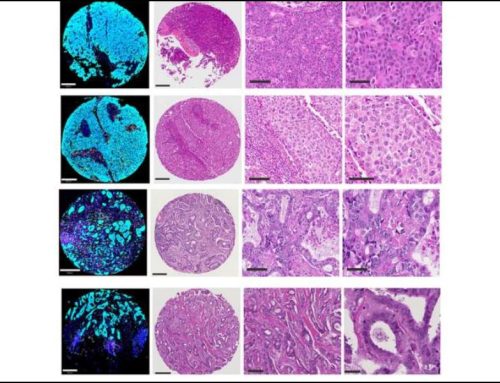
The research team conducted imaging experiments using various sample types and scales to validate the system. They imaged human tonsil tissue sections using both multiplexed RNA detection and protein immunofluorescence, achieving single-cell resolution and quantifying marker expression across thousands of cells. In mouse liver fibrosis models, the system performed large-area tissue scans with six-plex protein imaging, enabling automatic cell segmentation and the computation of spatial correlation patterns across different markers.
In addition, the PRISMS platform was able to localize individual mRNA transcripts in cultured fibroblasts and count them within segmented cell boundaries. The researchers also showed that the platform could perform super-resolution radial fluctuations (SRRF) imaging by acquiring repeated frames at each position.
“We consider the most striking result highlighting the versatility of PRISMS to be our ability to seamlessly switch between diverse sample formats and imaging modalities within a single unified pipeline, a flexibility we often find lacking in rigid commercial systems,” Zhang emphasized. “We believe this versatility was uniquely underscored by our integration of SRRF, which allowed us to acquire repeated frames and reconstruct super-resolved images, such as resolving mitochondrial features in stem cells, providing a level of imaging customization and resolution that is rarely available in standard ‘black box’ spatial omics solutions.”
The system also incorporates brightfield and darkfield imaging capabilities on the confocal platform using an LED matrix, enabling standard histological imaging (H&E, Masson’s trichrome) on the same tissue sections that are analyzed with fluorescence.
Potential Implications for Research and Clinical Labs
Zhang explained that affordable solutions like PRISMS could help democratize spatial omics technologies. Until now, advanced spatial analyses such as mapping tissue architecture in cancer and inflammatory, fibrotic, and neurodegenerative diseases have been largely confined to specialized research centers or industry partnerships.
“We envision PRISMS transforming the research landscape for smaller laboratories by drastically lowering the financial and technical threshold for entry, allowing them to compete in the rapidly evolving field of spatial biology,” Zhang stated. “By designing our software to function with standard, widely available microscopes and affordable automation tools, we allow labs to repurpose existing hardware rather than investing in million-dollar dedicated instruments.”
Future Directions
The platform requires some technical expertise in Python programming and microscopy, although the team has worked to minimize this barrier through documented notebooks and graphical user interfaces. Additionally, the system currently integrates only with Nikon microscopes and Cephla spinning disk confocals, limiting its applicability to laboratories with other equipment, although the open-source framework could be adapted to additional platforms.
Head-to-head comparisons between PRISMS and commercial systems using identical samples are needed to evaluate whether the cost savings justify potential trade-offs in performance metrics such as detection sensitivity, false positive rates, or throughput.
Zhang emphasized that the research team is focused on community adoption rather than commercial development.
“We have released our data, hardware designs, and pipeline code with the intent of providing a foundational, scalable method that other laboratories can optimize for their own diverse research applications,” he explained. “Our goal is to empower the wider scientific community to use and modify our tools to push the boundaries of spatial biology, enabling users to customize the workflow to their specific needs rather than waiting for us to release a commercial update.”
The study received financial support from the NSF CAREER and the National Institutes of Health.
References
- Zhang N, Fang Z, Kadakia P, et al. Modular, open-sourced multiplexing for democratizing spatial omics. Lab Chip. 2025;25(20):5379-5392. Published 2025 Oct 7. doi:10.1039/d5lc00286a