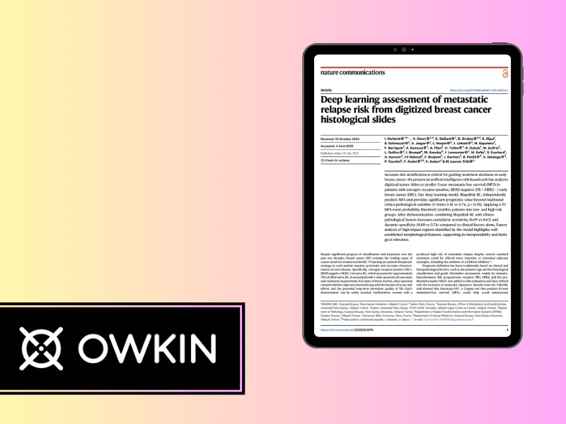
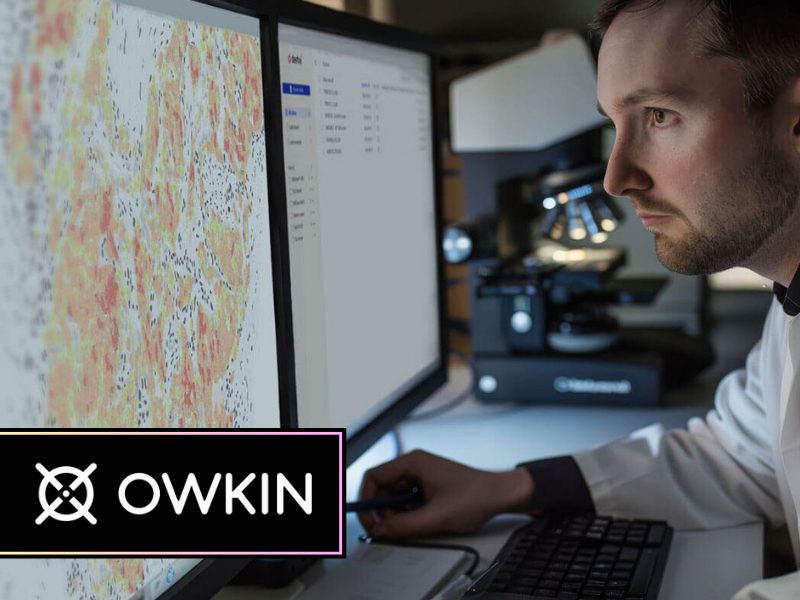
Owkin’s approach to integrating real-world hospital data with AI for digital pathology is unique. Unlike many companies that rely on publicly available data or cell models, Owkin built partnerships directly with hospitals to access clinical data while maintaining strict data privacy measures.They developed innovative solutions that allow hospitals to keep full control over their data by accessing it from behind hospital firewalls. This approach not only enhances data security and compliance with regulations like GDPR but also accelerates the adoption of AI in drug discovery, diagnostics, and patient care by leveraging real-world data at a large scale.
“The whole time I was doing my PhD, I could see the mirror between what we could see at the morphological level – that is a few microns of tissue – and what we could see at the molecular level, which is completely invisible to the naked eye. Being able to observe both at the same time was really exciting.” – Meriem Sefta
Conversation with Meriem Sefta
Owkin, Paris, France.
BIOSKETCH Meriem Sefta:
Meriem Sefta is the Chief Diagnostics Officer at Owkin, previously serving as Chief Data & Clinical Solutions Officer and Head of Partnerships. Before Owkin, Sefta was a Consultant at AEC Partners and a Research Engineer at Institut Curie. They also worked as a teaching assistant at Université Paris-Sud and a research assistant at MIT.
Sefta studied biology, chemistry, and computer science at Ecole polytechnique, earned a Master’s in biological engineering from MIT, and completed a PhD in computational biology at Institut Curie.
Interview with Jonathon Tunstall
Interview date – 10 November 2023
Publication date – 26 September 2024
JT – Ladies and gents, today my guest is Meriem Sefta from Owkin. Meriem, maybe you could tell us something about your background, your early career and how you came to be at Owkin and particularly interested in digital pathology.
MS – I took an interest in digital pathology before I joined Owkin. I was in grad school working on my PhD: specifically focused on retinoblastoma, which is a pediatric retinal cancer.
We were analyzing what I would say was the trend at the time, and still is, which was multimodal data on retinal cancer. So, we looked at genomic data, outcomes data, copy number alteration, methylation, and gene mutation data. We then started working with the pathology department at the Curie Hospital, where I was doing my PhD, to look at the digital pathology slides. I remember working with pathologists and seeing within the slides patterns that we would be seeing in the genomic data.
At this point, we were tying those two levels of information together, which was an eye-opener for me in realizing that the patterns that I could see at a bioinformatics molecular level, I could also to some extent sense from the pathology slides. To give you an example from the genomic data that I had,we had a few patient cases that were outliers and just didn’t look like the rest of the samples. When we looked at them unbiasedly with a pathologist, he looked at the slide and said, “huh, this one looks weird for a retinoblastoma. Typically, they don’t look like this. Let me just confirm the diagnosis,” He was that unsure because it was that much of an outlier in terms of what it looks like.
The whole time I was doing my PhD, I could see the mirror between what we could see at the morphological level – that is a few microns of tissue – and what we could see at the molecular level, which is completely invisible to the naked eye. Being able to observe both at the same time was really exciting.
I then joined a healthcare consulting firm after graduating, but in doing so, missed what was the more innovative space at the time. When I reached out to Owkin, they told me that they were working on artificial intelligence to better analyze these digitized pathology images. And I thought, well, yes, there’s definitely untapped information there.
So I thought, this is very exciting, because when you look at a pathology slide digitally, you can really see that there is a wealth of information in the image. To put that into context, a fully digitized pathology image is about two gigabytes of data, which is as large as a full-length feature film.
You could see that there was a wealth of information there, so for me it was a no-brainer with there being more information in these images than we were utilizing at the time. That’s why I joined Owkin. In the first years, we worked a lot on building research partnerships with pathology labs, and clinical researchers to start proof of concept on our hunch that there was probably more information in these pathology slides than what was currently being derived from them.
JT – Is this the point at which you formed the Owkin Data Alliance?
MS – Yes. I would say Owkin is pretty novel in that sense, from the beginning we didn’t work with only publicly available data or only molecular cell models like cell lines. I think the reason for that was that Thomas, our CEO and co-founder, was a clinician before joining Owkin, and with my background, we knew that going after hospital-level data was where there was a large wealth of untapped information.
With that in mind, we then had to get creative on how to do that, because I would say public-private partnerships between hospitals and startups or AI companies were still relatively rare at the time, especialy compared to the scale at which we wanted to do it.
And so, we had to find a way to roll out these academic research partnerships and get access to this data faster and at a broader scale than what had been done in the past. That’s where we innovated how we physically got access to the data. At the time, there was a lot of nervousness around data privacy and security, which still exists now. At that same time, GDPR had just started rolling out, so we built solutions that allowed us to access the hospital data from behind hospital’s firewalls. The result of that was that the data never left the governance, whether physical or virtual, of the hospital.
The major benefit of this was that the hospital never actually lost control of their data. And that was really important for building trust and setting up these collaborations relatively rapidly.
JT – Yeah, and I looked at your career history and the different phases you’ve had at Owkin, and I saw that in 2020, which is still quite recently, you changed role then and you set up this new department of data and clinical solutions. Maybe you could tell us something about that.
MS – Yeah, we continued building these partnerships for the first few years at Owkin, and then, across these research collaborations, we progressively started realizing that there were potentially lots of applications of this technology and the AI we had developed to better analyze these images, patient outcomes data and genomic data. We then had this major jump when we started building direct from R&D into the development of actual solutions that all used the same core assets: the data access and the AI tech.
We then developed them further and leveraged them for drug discovery. That means using the fundamental understanding that you have of your tissue or cancer, using the AI to better understand the mechanism behind a disease, and better identify potential drug targets for drug discovery.
We also developed this for drug development, which is a little further down the line. You have a potential drug candidate, but you really want to determine which patients are the best to recruit in your clinical trials. So, you need to know which biomarkers you need to measure to better predict response to treatment, and also to better stratify your patients for this new drug.
Similarly, you can also leverage the same data, the same tech applied to these images, and this genomic data to better stratify your patient pool for drug development. The third application was in the diagnostic space.
Basically, how can you then take these AI models and implement them into the real world? Using them as medical devices that pathologists or oncologists can leverage to stratify patients in the real world.
So, we developed applications across the entire value chain from discovery to diagnostics.
Interestingly, in doing this, we have come full circle from the initial data that we get from a care setting, right back into the care setting with these potential new drugs or new diagnostics.
JT – I think this is something that’s really fascinating because historically, if you look at this huge process of drug development, which let’s face it, can take 10 years from initial discovery of compounds to clinical trials.
Historically, those two facets have not been particularly linked. There were people who worked in discovery and they found, let’s say, a thousand potential compounds and they threw them over to toxicology and toxicology is based somewhere else where it’s done by another company. And maybe these people don’t even know each other.
And then eventually the toxicologists, they do their work and then they throw it over to be tested on animal models which is followed by clinical trials. Although these different groups are working toward the same goal, they don’t know each other, and that’s been the historical process within pharmaceutical companies.
And what you’re alluding to her, is putting some linkage using AI and digital pathology to link together the drug discovery process, correct?
MS – Yes, exactly.
JT – I think that’s a very powerful approach. I actually don’t know of another company that’s doing that right now. So, if we think about AI, the future potential of AI within drug discovery. What do you see as being the benefits of that? We have talked about linking it all together, but what are the real benefits that it brings to the process and maybe to the patient as well?
MS – Yes. I just want to emphasize that this is not as much my domain at Owkin as it is some of my colleagues, but for a lot of drug discovery up to today, I would say it has been carried out through animal models or cell line models. So, you model the disease, you identify potential targets, and then you go and test them in things that resemble humans more and more:
cell lines from human cancers, xenografts, and then eventually animal models and then in human trials. You start with a more theoretical approach, and then you progressively go towards models that resemble human diseases.
And here, the idea is to put the real-world patient data first and foremost in this process. So, base your discovery on real-world patient data and derive potential targets from the analysis of this data, The idea is if you do this from the beginning, you have things that are closer to reality. The hope is that this de-risks the entire drug discovery process, that historically can be very prone to error, where every step has a massive potential loss.
JT – Yeah. I think de-risking is a very powerful benefit that comes out of this. Developing drugs is very expensive, you’re talking a few billion dollars to bring a compound through to final clinical trial.
MS – Exactly! So, we’re not talking about a 200 percent optimization, but a 10, 15, 20 percent optimization. When you look at the bottom line of the whole process it’s a huge innovation. And for pharmas, it’s been very painful to access real-world data. It’s not that they don’t necessarily want to work with this type of data, and it’s not that they’re not convinced by this approach.
It’s just a very difficult process for them to have access to real-world data, clinical care data and, especially, multimodal data. So we stand out as an accelerator of this for our pharmaceutical partners in making this data access possible.
JT – Yeah, that’s great. Looking back on when I first started at Aperio, I sold digital slide scanners to pharmaceutical companies, and even back in 08, 09, AstraZeneca had 10 scanners, Novartis had 15 in different parts of the world. And that was really because of a shortage of pathologists in pharma and also they’ve got deep pockets, so they don’t mind spending a couple of 100,000 on a scanner here and there. And they use their international pathologists just by throwing the data around between them. But this is a completely different level of approach that we’re talking about now because this uses artificial intelligence to link together the drug discovery processes and I think that’s really fascinating.
MS – Yeah and we have a lot of collaborations with pharmas that are very strongly involved. For example, most of our data originates from our clinical network of partners, but also sometimes from pharma clinical trials. So, in one particular study or collaboration, we are compiling data from trials or studies that the pharma has sponsored and run them with real world data from the actual care setting. That’s where I think it becomes very rich in terms of approach.
JT – Yeah, great. I wanted just to change tack a little bit because I noticed you’ve got two products that are quite newly CE marked.
MS – Yes, that’s correct. We have one that’s called MSIntuit CRC, which is CE-marked. The other one, called RlapsRisk BC, was launched, but is not CE marked.
JT – There’s one for Colorectal cancer, right?
MS – Yes, that’s the MSIntuit CRC.
JT – Okay. So, perhaps you tell us a little about those individual products.
MS – MSIntuit CRC is the first of its kind almost globally in the sense that it’s a biomarker pre-screening solution that’s been regulatory approved and launched that uses digital pathology H&E slides.
What this means is, from the routine diagnostic H&E slide for colorectal cancer, we can pre-screen whether patients are likely to be carrying the MSI biomarker. Here, the idea is that the product is set up as a pre-screening step before the current gold standard test, which is IHC or PCR.
But what’s interesting is that the AI can identify patients who are not carrying this biomarker with very high sensitivity before we even run the molecular test. This phenotype biomarker is the microsatellite instability phenotype biomarker. It is very interesting at a clinical level because it is predictive of response to immunotherapy and patient outcome in response to different types of therapy in general. MSI-high patients in colorectal cancer have a different way of being cared for and very different outcomes from the rest of the colorectal cancer type patients.
At the clinical level, being able to identify these patients easily using this MSI phenotype is very important and what this solution allows you to do is reduce the testing burden because it pre-screens out all the patients that are negative for this biomarker. The outcome of the product is one of two things: either MSI negative or undetermined, in which case gold standard testing is what’s recommended. By using MSIntuit CRC you have ruled out a large proportion of patients directly from their diagnostic H&E slide without having to actually run the molecular testing, which is a significant advantage.
In terms of diagnostic speed, the H&E slide is routinely available. So this means that within half an hour of just running the AI, the doctor can get the response back as to whether or not this patient is carrying this biomarker. This is very important, especially in oncology, where traditional molecular biomarker testing methods can be so long that clinicians will just go ahead and start treating without actually getting the test results.
Also, with the number of cancer patients growing, as well as the number of biomarkers to test for, the burden on healthcare systems is becoming larger, creating longer backlogs and increasing the testing burden for pathology labs. So, this time-saving argument is critical when it comes to patient care.
The second argument for using MSIntuit CRC is that once it is run you don’t actually even need to be running the molecular tests in a lot of cases. This spares samples, time, materials, and all those other things that would have been necessities. Interestingly, for some biomarkers and in some geographies, the MSI testing is deemed too expensive, too complicated, or too long for it to even be systematically indicated, so they won’t even do it because of these hurdles. For example, in France, it’s not so much the case that the testing isn’t done, but in other geographies, these biomarkers aren’t even systematically tested for. So, if we can use AI we can speed up the process by ruling out a bunch of patients and focusing on specific patients that are very highly likely to be carrying this biomarker.
The MSI phenotype is present in 15 percent of the population. So, you need to test 100 to find 15. In the latest version, we rule out almost 50 percent of the population. This means that you test 50 to find 15. We’re now rolling out the concept of biomarker pre-screen from digital pathology slides to other use cases. MSIntuit CRC is, I would say, the first in a series of products that all share this same backbone.
We’re extending this MSI solution to other indications and also extending this concept of biomarker prescreening to other biomarkers. So, in essence, we’re overall reducing the testing burden because, again, we’re always using the same diagnostic H&E slide.
What you can imagine in the future is that from the same slide, you can have information such as ‘highly unlikely to be carrying MSI’, ‘highly likely to be carrying this mutation’, ‘highly likely to be part of this subgroup’, etc. AI has the promise of becoming a very strong complementary pre-testing step to more expensive and more difficult to roll out molecular testing.
JT – And of course you are, you are avoiding the need for multiple layers of IHC, for example, because you’re diagnosing from an H&E and it’s really a personalized medicine approach using AI, the AI becomes an integral part of the diagnosis. And you can see that in the future, AI could detect things that the human eye couldn’t detect, for example, you could actually find AI biomarkers.
I planned to ask you this question, but this is a very crowded field, if we look at digital pathology, and we look at algorithms, image analysis, artificial intelligence, and applying that to digital slides. It’s a very crowded market. We were at a conference in Budapest back in the spring, and I think there were 18 companies producing some kind of algorithm in digital pathology.
How does Owkin stand out from the crowd in this market and stay relevant?
MS – I would say two things. One, a lot of these companies focus on what I would call pathologist support tools. So solutions that do what pathologists currently do today, either automatically or through AI. For example, automatic tumor segmentation or automatic tumor grading.
Here, through this MSI product, as well as RlapsRisk BC, which predicts risk of patient relapse in breast cancer, we’re not just supporting the pathologist, we’re going beyond what the pathologist eye can do. We’re pre-screening for biomarkers that would otherwise need molecular testing to be identifiable.
If we’re in the case of RlapsRisk BC, we’re even predicting the risk of patient relapse directly from the digital pathology side. That’s also something that, until today, was only done through molecular panels and expensive testing. So, in terms of ultimate added value and unmet medical need, we’re answering more difficult and higher unmet medical needed use case. I think that’s one very strong differentiator.
Another aspect is around advancement of the actual data science algorithm development. This is just the tip of the iceberg.
In terms of costs and investment, we’ll invest maybe 15 percent of our resources on data science, 30 percent on building a network of data labs and getting diverse data sets as much as possible to be able to train, validating and testing our models, and then a large part on engineering and regulatory to really make them into certifiable, safe and efficient products that can be used in a real world setting.
A lot of companies don’t have these capabilities yet, and I think we do. We are much more advanced in that field, as well. Especially the industrialization process. There have been some scientific publications on the theoretical feasibility of some of these use cases at a data science level, but not at a productized regulatory-approved level.
There are a lot less players on the market if you’re looking at how much AI is marketed and what’s pitched today. A lot of what’s pitched and marketed today are not mature products, it’s more product prototypes. I think in the ability to roll them out into products, go to market, and re-leverage at a clinical level, this network of data partners creates a strong differentiator for us, because then we re-deploy these solutions in the same labs as the ones that we collaborated with for the product development. It’s like end-to-end of what we’ve been doing.
JT – As you said, the product maturity, I think that’s a really important point as well. Again, quite a general question, but we talked then about the pathologist. We’ve been talking a lot about AI. How’s this going to change the world of pathologists? Because that is going to change dramatically?
What are your thoughts on that? The way the pathologist works in 10 to 15 years time.
MS – So the good news is pathologists are very excited about this, for several reasons I think. One, they see that something’s got to give, like the number of tests that they need to run is becoming higher.
The cost burden is also becoming higher. The number of biomarkers out there is increasing. Precision medicine is becoming more and more of a reality, but then this is putting extreme pressure on pathology labs to be able to run these diagnostics faster to be able to basically identify more and more biomarkers.
And anything that can allow them to pre-screen to directly identify a whole bunch of things directly from the diagnostic slides, this not only accelerates their work, it potentiates how much they can provide and in faster time to clinicians in terms of information on the patients.
A lot of the advances in precision diagnostics in the past years have been around molecular biomarkers. For pathologists, this is also a way to have solutions for them to stay like a strong player in this game of being able to provide relevant information from these pathology slides.
They’re very excited about it, and we definitely have very strong support. They do know that this is going to disrupt the way that they do things, and I think what’s exciting to see is that in all our target markets, we’re seeing digital pathology associations asking: how is this going to impact what we do?
How do we need to start preparing for it? How do we need to change the way we train pathologists to be better trained for this digital transformation?
It’s the same transformation that happened for radiology when it went progressively digital and then they started progressively adopting AI solutions for diagnostics. And here, I would say, it’s the same thing, just a lot faster because the AI solutions are arriving on the market at the same time the labs are going digital. So, it’s just compressed in terms of timeline, and you have this senior crowd of pathologists who are scrambling to not only adopt this digital transformation, but also get ready for the future and the digital pathology diagnostics that go alongside it in a very quick timeline.
So I think it’s a pretty exciting time for me in general.
JT – I agree. You mentioned that about training. I think that’s a really important aspect for the future because if you think about current laboratories, which are based on microscopy, it’s hard to see how that can continue because there are young people coming into the profession who are trained digitally.
A lot of new young pathologists have been trained on digital slides and I’ve heard stories of people not knowing how to actually diagnose from a microscope. So, if you’re a laboratory that only has microscopes, how will you recruit into those labs in the future? That’s also a very interesting point, which I think will push the market more towards digital.
Or maybe we see two levels of service so there is a basic service that uses microscopy and it’s traditional, and you pay less money for that. And then you have a higher level of analysis at a higher price.
MS – What we’ve seen, at least in France, is an issue of talent retention. So if you’re a pathology lab, and you’re working with classical microscope, and don’t have AI or digital solutions, it’s going to be a lot harder for you to compete and hire key pathologist talent, especially for the younger generation, than a lab that’s fully digital and allows a pathologist to work from home. They have cutting edge AI solutions in place and there’s a strong push for all labs to catch up. In the beginning, I would say about four or five years ago, the first labs that went digital were clearly very early adopters and early adopter profiles. Now it’s accelerating and that’s where it’s very exciting, because you see everybody getting into gear to do this and everybody realizing that if they don’t catch up, they’re going to be the ones left behind.
And this switch happened, I would say, in the last one or two years. We started seeing strong acceleration. The same digital pathology conferences that some people had been going to for years, suddenly became very big.The venues were fully booked, and they had to change the size of the venues in order to welcome more and more people, because this is no longer perceived as a gadget.
JT – Just as a concluding remark, the future of pathology and the pathologist is very bright, both of them. This is not a technology that will make pathologists redundant or replace them.
In fact, they will still be signing off their cases and then they will have a host of new tools at their fingertips which will give a greater depth and clarity to the diagnosis, which at the end of the day benefits the patients, right?
MS – Exactly.
Share this article

Follow us
Get Pathology News Delivered to Your Inbox