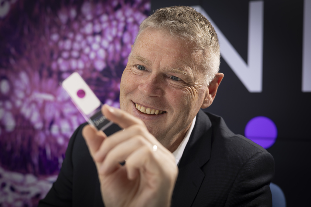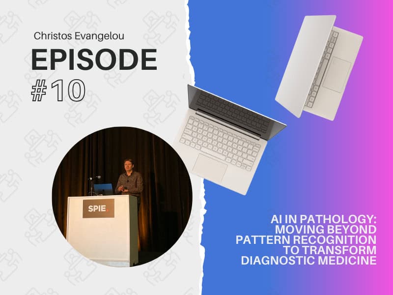
The future of computational pathology extends far beyond automated pattern recognition, according to Geert J. S. Litjens, PhD, who delivered a compelling keynote address.[1] Speaking to a packed audience, Litjens outlined how AI is revolutionizing diagnostic medicine through multimodal integration and scientific discovery.
“Most of our work in computational pathology has focused on mimicking what pathologists already do,” said Litjens. “While this is useful for improving efficiency and supporting physicians, the real potential lies in using AI for scientific discovery and driving fundamental oncological research.”
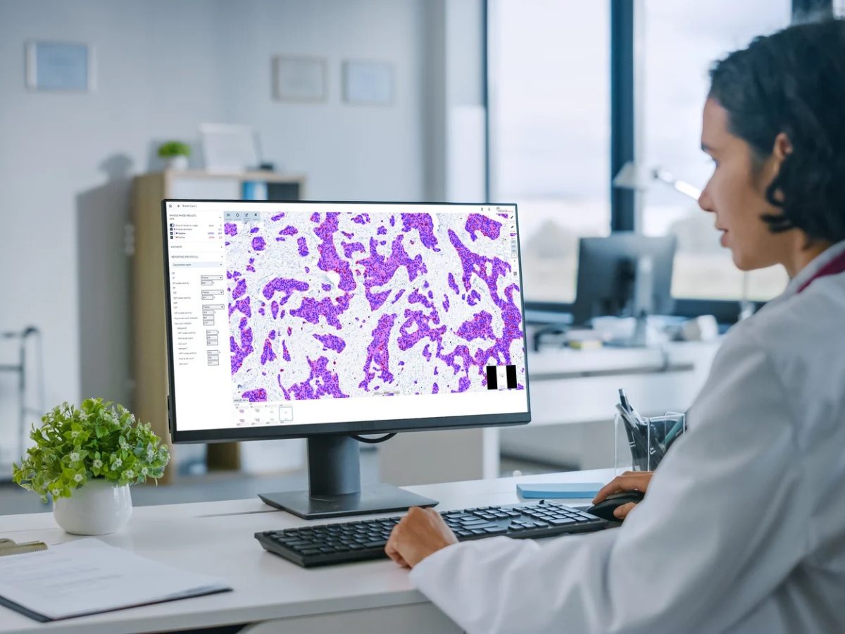
The field has come a long way since its early days. Litjens recounted how the 2010 volcanic eruption in Iceland unexpectedly advanced computational pathology when it stranded researchers in the Netherlands, leading to crucial collaborations. This is how Litjens started his PhD research on computer-aided detection of prostate cancer using MRI. The real transformation began with the application of deep learning to WSIs in 2013–2014.
Litjens highlighted recent advances in the direct prediction of patient outcomes from tissue images. In a 2023 study on prostate cancer, his team developed models that could predict the time to biochemical recurrence from tissue microarray samples. When combined with traditional pathologist assessments, these AI predictions provided complementary prognostic information. “The algorithms aren’t just replicating what pathologists see — they’re finding different patterns,” Litjens noted. “This is where computational pathology can discover new morphological biomarkers to improve patient prognosis assessment and potentially guide treatment selection.”
Recent advances in foundation models, which are pre-trained on vast datasets of images, have enabled the application of AI-aided systems for the detection of rare cancers with limited samples. However, Litjens cautioned against viewing these as a complete solution, noting that current foundation models still struggle with center-specific variations and image artifacts.
One promising direction is the development of “streaming stochastic gradient descent,” which allows end-to-end training with WSIs while maintaining manageable memory requirements. This approach has shown better generalization to external datasets compared to traditional methods.
Looking ahead, Litjens emphasized the importance of moving beyond single-modality AI to consider the entire patient journey. “We need to think less about being in pathology or radiology and more about integrating information across the patient’s diagnostic and treatment pathway,” he said. This integration is particularly relevant for diseases such as prostate cancer, where decisions are informed by multiple data streams, including blood work, imaging, pathology, and molecular diagnostics. However, Litjens disagrees with approaches that simply ‘freeze’ individual AI models and combine their outputs.
“In the clinic, we don’t train specialists and then freeze them — they work as teams and learn from each other,” he explained. “If a pathologist tells a radiologist they are getting too many benign cases with inflammation, the radiologist adjusts their guidelines. Our AI systems should have similar flexibility to learn and adapt.”
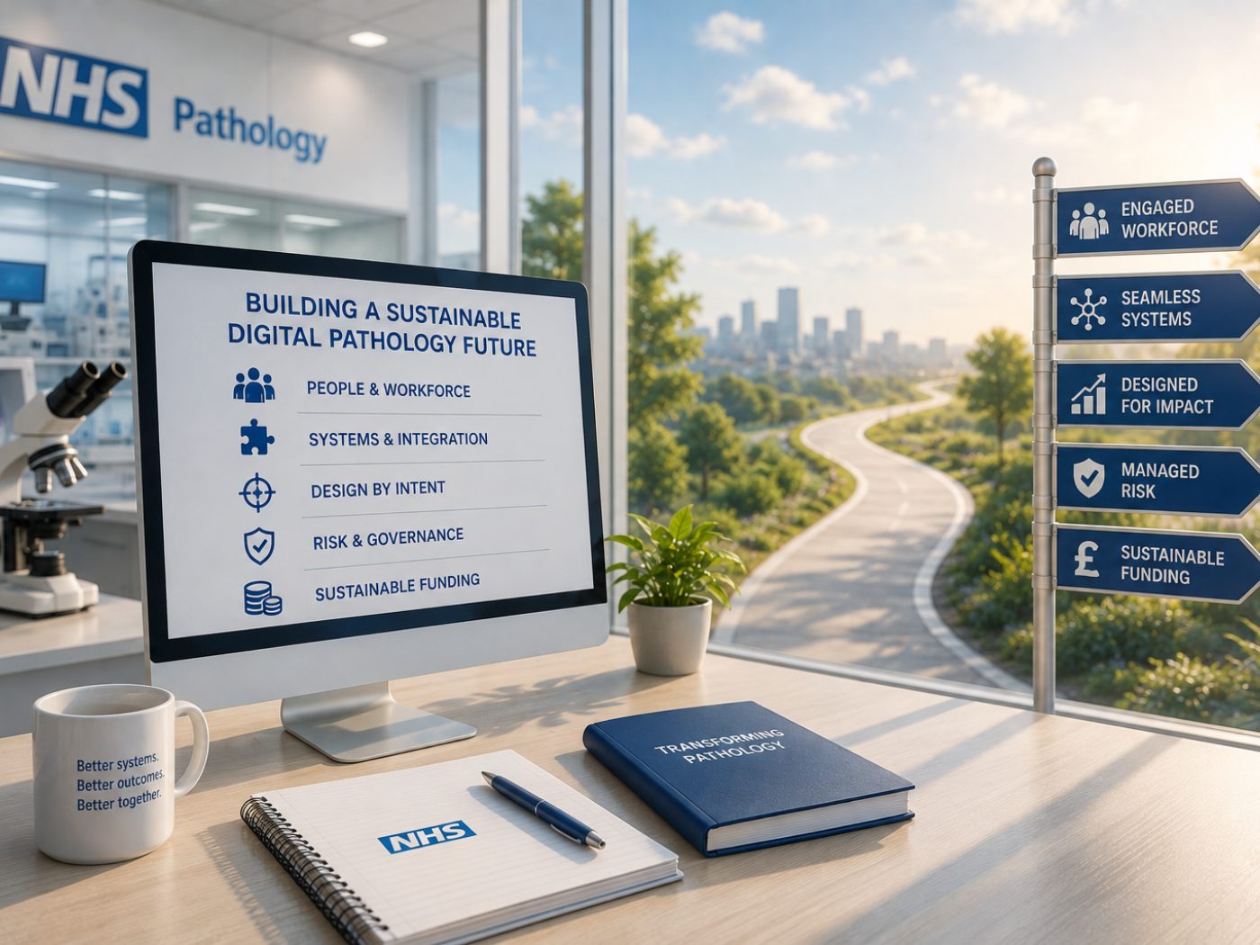
When asked about implementation challenges, Litjens acknowledged that widespread adoption faces hurdles beyond technical validation. “This isn’t a quality question — there are several high-quality products on the market,” he noted in a post-presentation interview. “The issue is that you can only use it effectively with a fully digital workflow and high-quality AI integrations. In Europe, there’s also the question of who pays for these algorithms, as hospital budgets are already stretched thin.”
Litjens expressed concerns about the potential misuse of AI in healthcare. “My nightmare scenario would be health insurers mandating AI to decide whether a patient gets specific treatments, bypassing both physician and patient,” he cautioned. “This is why it’s essential for physicians to participate in AI development and implementation, so we deploy it ethically while dealing with the reality of increasing healthcare cost and aging of the general population.”
To accelerate progress, Litjens advocates for more open data sharing and scientific benchmarking competitions. His team organized multiple challenges that have helped drive rapid improvements in various applications, from lymph node metastasis detection to Gleason grading.
Litjens also emphasized the crucial role of pathologists in driving further progress in computational tools. “For AI developers, it is absolutely essential to work closely with doctors,” he said. “They contribute meaningful challenges to tackle, have deep knowledge of the disease and underlying data, and are the end-users who ensure AI is integrated effectively.”Geert J. S. Litjens, PhD, professor of AI for analysis of medical images in radiology and pathology at Radboud University Medical Center, Netherlands.
[1] Geert J. S. Litjens, The future of diagnostics: the role of computational pathology in tomorrow’s medicine. Presented at SPIE 2025 Digital and Computational Pathology conference, February 19, 2025; San Diego, CA.
No audio available for this article yet.
No quiz available for this article yet.