by Christos Evangelou, MSc, PhD – Medical Writer and Editor
In a recent validation study, researchers at the University of Pittsburgh evaluated the performance of the Hologic Genius Digital Diagnostics System (GDDS), a new artificial intelligence (AI)-powered digital diagnostics solution, in analyzing ThinPrep Pap tests. The sensitivity for the detection of high-grade cervical lesions with the digital diagnostics system was 100% when considering Pap results of atypical squamous cells and above, with strong agreement between the pathologists reviewing the cases.1
“Revolutionary AI technology has the potential to completely revamp gynecologic cytopathology practice,” said Dr. Lakshmi Harinath, Assistant Professor of Pathology at the University of Pittsburgh and the lead author of the study. “Utilization of these innovative systems can mitigate workflow problems faced by laboratories due to staffing shortages, thereby reducing workload and providing relief from monotony and health issues endured by cytologists while screening Pap slides over a long period of time.”
The report was published in Cancer Cytopathology.
Need for Innovation in Cervical Screening
Cervical cancer is the fourth most common cancer among women globally, with early detection through screening being crucial for successful treatment.1 The introduction of the Papanicolaou (Pap) test has dramatically reduced cervical cancer mortality through the detection of precancerous lesions, such as high-grade squamous intraepithelial lesions (HSIL). However, this screening process is labor-intensive and imposes a significant burden on cytotechnologists and pathologists. With diagnostic laboratories facing increasing staffing shortages, there is a need for automated solutions that can maintain or improve diagnostic accuracy while increasing efficiency.
The Technology
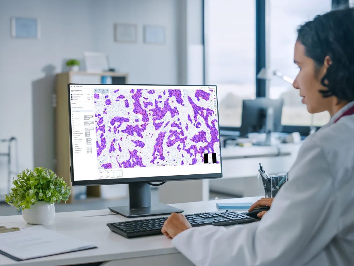
GDDS represents a significant advancement in digital cytology and has been recently cleared by the FDA for commercial use in clinical practice.1 The system combines AI algorithms with volumetric imaging technology to scan ThinPrep Pap test slides across multiple focal planes and analyze the cells. The system can scan entire slides and process up to 14 focal planes simultaneously, creating a comprehensive view of all cells present. After analyzing tens of thousands of cells, the AI algorithm selects 30–60 of the most diagnostically relevant cells and presents them to cytopathologists in an organized gallery format.1
The system categorizes these cells based on various characteristics, including nuclear-to-cytoplasm ratio and cell morphology, helping pathologists focus on the most suspicious cells.1 This approach aims to streamline the screening process while ensuring that no significant abnormalities are missed.
Study Design and Methodology
“It is imperative to confirm that the new system will meet the high quality standards of testing, providing accurate diagnoses essential for patient safety,” said Dr. Harinath. “Hence, we wanted to study the sensitivity of the GDDS for HSIL diagnosis and to make sure that this process is replicable amongst different pathologists interacting with the system.”
The researchers conducted a validation study involving 890 ThinPrep Pap tests, with a sub-study focusing on 183 cases that had been diagnosed as HSIL and confirmed through biopsy.1 Three experienced cytopathologists independently reviewed these cases using the GDDS, with support from cytotechnologists who performed initial screening. The pathologists were provided only with patient ages and were blinded to clinical history, HPV status, and original diagnoses.1 The study participants received two days of training on the system before beginning their evaluations.
Key Findings
The GDDS demonstrated high sensitivity rates for detecting high-grade cervical lesions. When considering all abnormal results (ASC-US and above), the system achieved 100% sensitivity for detecting cervical intraepithelial neoplasia 2 and above (CIN2+) across all three pathologists.1 For high-grade abnormalities (ASC-H and above), sensitivity ranged from 84.7% to 92.9% among the pathologists.
Dr. Harinath noted that these findings were very encouraging.
“All of the cases with a squamous cell abnormality were interpreted as ASC-US and above, with the majority of HSIL cases being accurately diagnosed as high-grade lesions by all three participating cytopathologists,” she said.
None of the HSIL cases were misclassified as normal, although some were categorized as lower-grade abnormalities. The percentage of cases classified as low-grade squamous intraepithelial lesions ranged from 5.5% to 11.5% among the three pathologists, and atypical squamous cells of unknown significance (ASC-US) classifications ranged from 1.6% to 3.8%.1
The study also demonstrated a strong agreement between pathologists, with a Kendall W coefficient of 0.722. According to Dr. Harinath, this high agreement rate was
“a pleasant and surprising result because the pathologists interacted with the system with minimal training and did not have the patients’ prior history or HPV status at the time of interpretation.”
Implications and Implementation Challenges
According to Dr. Harinath, the study findings suggest that the implementation of AI-powered digital diagnostic systems can help address the growing shortage of skilled cytotechnologists and improve diagnostic accuracy and efficiency in cervical cancer screening.
“AI-assisted technologies like the Genius system can help standardize diagnostic interpretation across lab systems with the possibility of remote expert consultation aiding in accurate diagnoses,” Dr. Harinath said. “As more and more labs adopt these systems, widespread collaboration for clinical as well as research purposes is a definite and exciting possibility.”
However, the application of GDDS in this validation study presented unique challenges that may indicate potential difficulties when transitioning to digital cytopathology in real-world settings.
“The main challenge was to get the team to become comfortable with the system in such a short time and to be able to transcribe their diagnostic skills from the microscope to the digital milieu,” Dr. Harinath explained. “The subtle changes in morphologic diagnosis seen when 3D images are presented as 2D pictures do take a little time to get used to.”
Looking Ahead
Dr. Harinath emphasized that additional studies are needed to fully understand the system’s performance across all diagnostic categories and its impact on the laboratory workflow. Future research should also examine patient outcomes and the performance of the system in routine clinical practice with fully trained users.
Dr. Harinath added that the successful integration of AI systems in routine clinical practice will depend on several key factors.
“The drive and desire for innovation by the relevant stakeholders, the evolution of newer prototypes, and increasing awareness of the availability and benefits of utilizing such systems are important considerations,” Dr. Harinath said. “Adoption of an open and ‘cautiously optimistic’ attitude with ‘evidence-based decision making’ is the key to successful integration of such systems in clinical practice.”
Dr. Harinath sees particular promise in the potential of the technology to expand access to expertise.
“New avenues for remote clinical sign-out, education, and research collaborations are compelling reasons for utilization of these novel technologies.”
- Harinath L, Elishaev E, Ye Y, et al. Analysis of the sensitivity of high-grade squamous intraepithelial lesion Pap diagnosis and interobserver variability with the Hologic Genius Digital Diagnostics System. Cancer Cytopathol. Published online November 5, 2024. doi:10.1002/cncy.22918
No audio available for this article yet.
No quiz available for this article yet.