by Christos Evangelou, MSc, PhD – Medical Writer and Editor
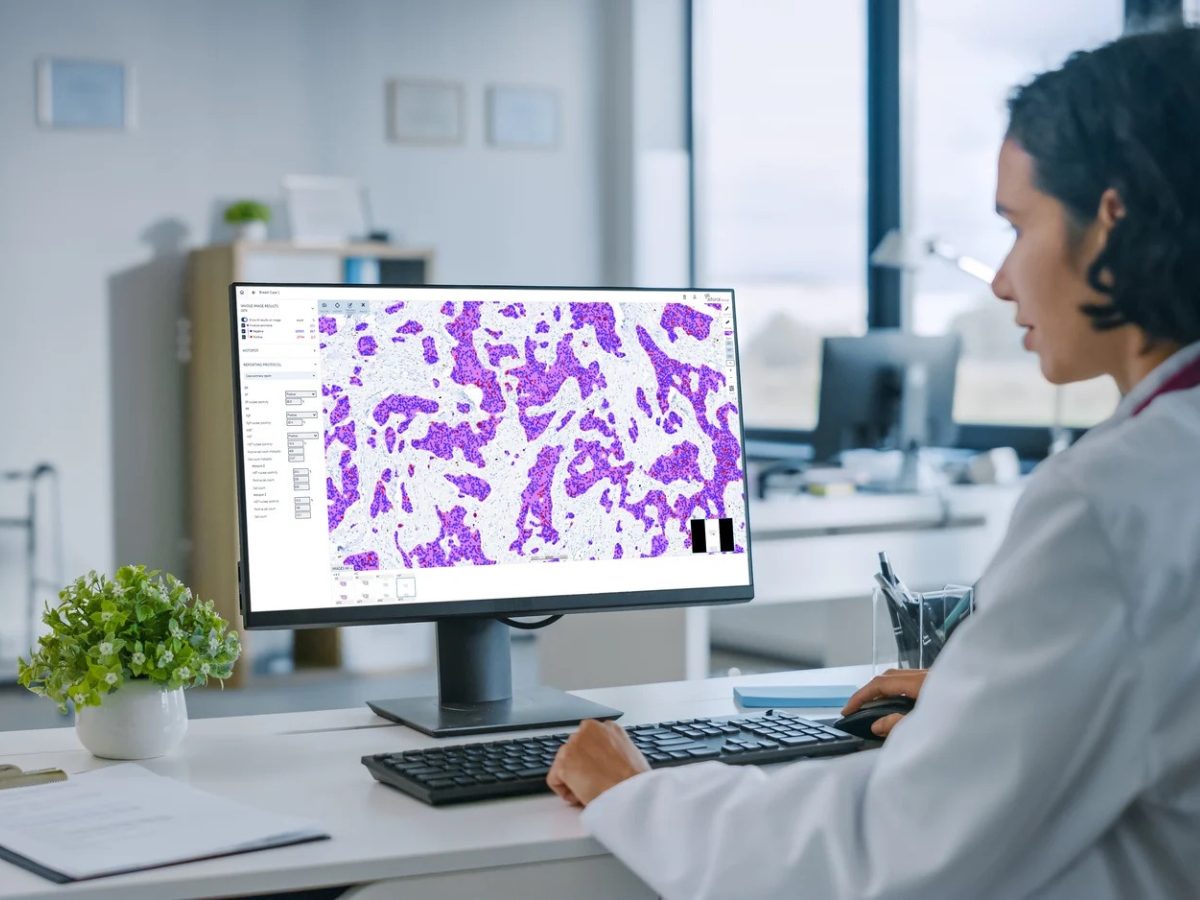
Researchers at the University of New South Wales and St. George Hospital in Sydney, Australia, developed a new artificial intelligence (AI) framework that combines innovative deep learning techniques to create a more reliable and efficient diagnostic tool for pathologists. The system, termed HistopathAI, achieved high accuracy in analyzing microscopic tissue images across multiple types of cancer, potentially reducing diagnostic errors and improving patient outcomes. According to the authors, this new framework could enhance workflow efficiency and diagnostic standards and facilitate multicenter collaborations by supporting the shift to digital pathology.
“HistopathAI consistently achieved state-of-the-art classification accuracy across all evaluated datasets, outperforming baseline models and previously reported methods,” noted Md Mamunur Rahaman, PhD candidate at the University of New South Wales and the lead author of the study. “The system delivered near-perfect accuracy on the in-house dataset, demonstrating remarkable robustness and precision. In challenging multiclass scenarios, HistopathAI also substantially surpassed other approaches, offering both higher accuracy and greater stability.”
The report was published in the Journal of Advanced Research.
The Critical Need for Better Diagnostic Tools
Cancer diagnosis through histopathology remains the gold standard for confirming cancer presence and type. However, manual microscopic analysis of tissues by pathologists can be subjective and prone to variability, with some studies showing accuracy rates as low as 57% for certain cancer subtypes.1 This variability can lead to delayed or incorrect diagnoses, potentially affecting treatment outcomes. This challenge is further complicated by the increasing global cancer burden, shortage of pathologists in many regions, and complexity of cancer diagnosis.
“Our motivation for this study arose from the limitations of traditional histopathological workflows, which often face inter- and intra-observer variability and struggle to generalize across diverse patient populations and institutions,” explained Rahaman. “As the complexity of digital pathology data grows, and as more laboratories transition to fully digitized slides, we increasingly need accurate, consistent, and scalable diagnostic tools.”
A New Approach to Histopathological Image Analysis
The researchers developed HistopathAI, the architecture of which combines two powerful deep learning models, EfficientNetB3 and ResNet50, with an advanced learning strategy called supervised contrastive learning (SCL).1 What sets HistopathAI apart is its hybrid deep feature fusion technique, which merges information from both models to create a more comprehensive understanding of tissue images than traditional single-model approaches.
“Traditional methods like cross-entropy loss directly optimize classification accuracy, which can lead to skewed or less discriminative feature representations, especially in imbalanced datasets,” Rahaman said. “SCL, by contrast, refines the embedding space using a temperature-scaled cosine similarity measure. It ensures that samples of the same class are tightly grouped together while pushing apart samples of different classes.”
The system uses a two-stage training process: first, learning to recognize important features in tissue images, then refining its ability to classify these features into specific cancer types.1 Rahaman explained that this approach mirrors how human pathologists develop their expertise but with the added advantage of computational precision and consistency. The system can simultaneously analyze multiple levels of tissue features, from cellular structures to broader tissue patterns, providing a more complete diagnostic picture.
Rahaman noted, however, that the expanded feature space elevated the risk of overfitting, particularly in scenarios with limited training data. He added that fusing features from EfficientNetB3 and ResNet50 demanded careful alignment to ensure the resulting combined features were meaningful and compatible.
“We applied global average pooling layers to extract comparable, compact feature vectors, introduced dropout and other regularization methods to mitigate overfitting, and employed stratified cross-validation to optimize hyperparameters and evaluate model stability,” Rahaman said.
Framework Validation and Potential Implications
The team tested HistopathAI using eight different datasets, including seven publicly available collections and one private dataset, encompassing various types of cancer, including breast, colorectal, and gastric cancers.1
HistopathAI exhibited promising performance across multiple metrics. The system achieved over 99% accuracy in several datasets, outperforming existing methods.1 Notably, the framework demonstrated superior performance in both binary (cancer vs. non-cancer) and more complex multiclass classifications, such as the 7-class Breast Carcinoma Subtyping dataset, where traditional methods often struggle.
HistopathAI maintained high performance even with imbalanced datasets, a common challenge in medical imaging where certain cancer types may be underrepresented.1 The system also maintained high accuracy across different types of cancer and varying image quality, suggesting its real-world applicability.
According to Rahaman, these findings highlight the potential impact of HistopathAI on clinical practice.
“HistopathAI can be readily integrated into digital pathology workflows, serving as a pre-screening or decision-support tool to help pathologists prioritize critical cases and streamline their diagnostic processes,” Rahaman explained. He added that “its modular design and two-stage training approach allow for efficient fine-tuning, enabling it to adapt to new datasets and institutional protocols with minimal additional labeled data.”
Overcoming Challenges and Future Directions
Despite the promising performance of HistopathAI, the authors foresee hurdles to its clinical implementation.
“While HistopathAI delivers robust performance, the ‘black-box’ nature of deep learning models can make it challenging for clinicians to trust and fully understand its decision-making process,” Rahaman noted. “Future efforts will focus on incorporating explainable AI techniques, such as saliency maps or attention-based methods, to highlight the regions of interest and provide insights into the model’s reasoning.”
Rahaman revealed the team’s current efforts to further improve model robustness and address issues such as dataset imbalance. These efforts include using generative adversarial networks or diffusion models to create synthetic samples, developing methods for domain adaptation and federated learning, and combining histopathology images with genomic, proteomic, and clinical data.
The team is particularly focused on making the technology more accessible.
“We are working on streamlining architectures for deployment in resource-constrained settings, ensuring that advanced diagnostics are accessible globally,” Rahaman explained.
This commitment to accessibility could help address healthcare disparities in regions with limited pathology resources.
The study received financial support from MTPConnect and MRFF Australia through a Researcher Exchange and Development in Industry Fellowship to Ewan K. A. Millar.
References
- Rahaman MM, Millar EKA, Meijering E. Generalized deep learning for histopathology image classification using supervised contrastive learning. J Adv Res. Published online November 16, 2024. doi:10.1016/j.jare.2024.11.013
No audio available for this article yet.
No quiz available for this article yet.