by Christos Evangelou, MSc, PhD – Medical Writer and Editor
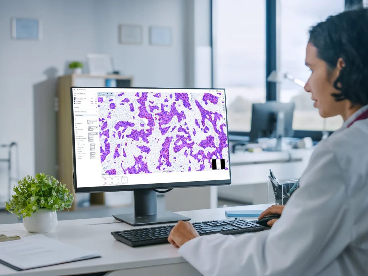
Until recently, the anatomic pathology laboratory had remained largely unchanged. However, over the last few years, there has been an exponential increase in the adoption of digital pathology solutions. Keith Wharton Jr., MD PhD, a pathologist and Global Medical Affairs Lead at Roche Diagnostics Solutions, led a team that used the Roche Digital Pathology Dx Whole Slide Imaging (WSI) system to analyze over 2,000 clinical cases, involving 16 pathologist readers across multiple sites. The study showed digital pathology to be noninferior to traditional microscopy for primary surgical pathology diagnosis, with a difference in accuracy of -0.61%. The Roche Digital Pathology Dx system received FDA 510(k) clearance in June 2024.
In an interview with Pathology News, Dr. Wharton discusses Roche’s recent digital pathology validation study and what it means for the future of pathology. The study findings were published in the American Journal of Clinical Pathology.
The Start of Roche’s Journey in Digital Pathology
“Roche has a long-term commitment to bring digital pathology to tissue diagnostics,” Dr. Wharton said, noting the company’s acquisition of digital pathology pioneer Bioimagene in 2010. “But for the USA market, FDA clearance for primary diagnosis is the ‘price of admission’ to be taken seriously by diagnostic laboratory customers.”
Although the system performance studies were planned before the pandemic, COVID-19 created an increased need for remote diagnostic capabilities.
“There were a lot of questions about whether our study could be completed, given the need for pathologist readers and a critical shortage of onsite healthcare workers,” Dr. Wharton said. “We and our external collaborators made the best of a tough situation to push forward.”
Assessing the System’s Accuracy
Unlike previous studies that used fewer adjudicators, The Roche team employed 20 pathologist adjudicators to evaluate diagnostic concordance. This larger pool enabled the researchers to accelerate study completion and showed that the length of reference diagnoses has a significant impact on disagreement rates, regardless of whether the reading pathologists used digital or manual microscopy.
“During the planning stages, due to high clinical demands, all four of our external clinical sites did not have time to shorten the input diagnoses from the medical record,” Dr. Wharton explained. Some reference diagnoses contained thousands of characters and entire pathology reports rather than concise diagnostic statements. “This had the effect of ‘kicking the can down the road’ for the adjudicators to sort out.”
The researchers found that when adjudicators compared microscopic and digital reader diagnoses directly to each other, rather than each of them to lengthy reference diagnoses, the disagreement rate dropped to 4.77%. Using the reference diagnosis as a “tiebreaker” further reduced the digital pathology disagreement rate to 2.97%.
“The adjudication process itself is ultimately subjective,” Dr. Wharton said. “It stands to reason that the more adjudicators you have, the more variation in their performance you will see, which is exactly what our data demonstrated.”
The Convergence of Technologies
Dr. Wharton commented on how similar all FDA-cleared digital pathology systems have performed compared to the first system clearance in 2017. Their study joins five (now six) other WSI systems using novel scanners in showing consistent results across different manufacturers and technologies, all achieving the noninferiority threshold with differences of approximately 1% compared to manual microscopy.
“Digital pathology scanners have been around for a couple of decades, and while they each use slightly different technology, what the aggregate results tell us is that as long as you’re using a system that produces an image above a certain quality threshold, they all work fine for primary diagnosis,” he stated.
Dr. Wharton emphasized that, although all the tested scanners are largely equivalent for primary diagnosis, they all produce images that look slightly different, even from the same slide, so there are aesthetic preferences at play, and these differences could also drive unexpected changes in performance of image analysis algorithms that use artificial intelligence (AI).
The Effect of Magnification on Diagnosis
The study showed that only 0.18% of over 3000 slides required 40× magnification for diagnosis, supporting the adequacy of 20× scanning for most cases. This finding has implications for laboratory efficiency.
“A 40× image file with no compression or other file manipulations is approximately four times larger than a 20× image,” Dr. Wharton noted. “If you can make adequate diagnoses from a 20× slide, there’s no reason to do a 40× scan, at least for primary diagnosis.”
The finding offers laboratories confidence that 20× scanning will cover the vast majority of cases while providing advantages in workflow and storage costs.
Potential Challenges
The study identified some expected organ-specific challenges in tissue analysis using digital pathology, particularly with breast in situ carcinoma cases, where digital reads showed a slight tendency toward “undercalling” compared to manual microscopy. However, Dr. Wharton cautioned against overinterpreting these findings.
“None of the organ groups and sample type subgroups were adequately powered to make definitive conclusions, especially in the post hoc setting,” he emphasized. “The percentage of breast in situ carcinoma case types with discrepancy in digital mode was small, less than 10% of an underpowered subcohort size.”
He added that the distinction between atypical ductal hyperplasia and in situ carcinoma can be very challenging in any modality.
“There’s some plausibility for a 20× WSI to rarely lead to undercalling, since the diagnostic criteria rely partly on fine examination of nuclear chromatin features that may be more thoroughly examined on a microscope where one can focus up and down at high magnification,” Dr. Wharton said.
Dr. Wharton advises pathologists to carefully assess digital image quality, and if the quality is insufficient to make a confident call, they should obtain a rescan, a higher magnification scan, or use their microscope.
Rethinking Regulatory Requirements
With seven FDA cleared systems with novel scanners now showing very similar performance, Dr. Wharton believes the time is ripe for regulatory pathways to evolve.
“We suggest in the paper that usability studies and analytical evidence of image quality might be sufficient to ensure safety and effectiveness of WSI systems for primary diagnosis — just as they are for the microscopes that pathologists have used for primary diagnosis for nearly two centuries.”
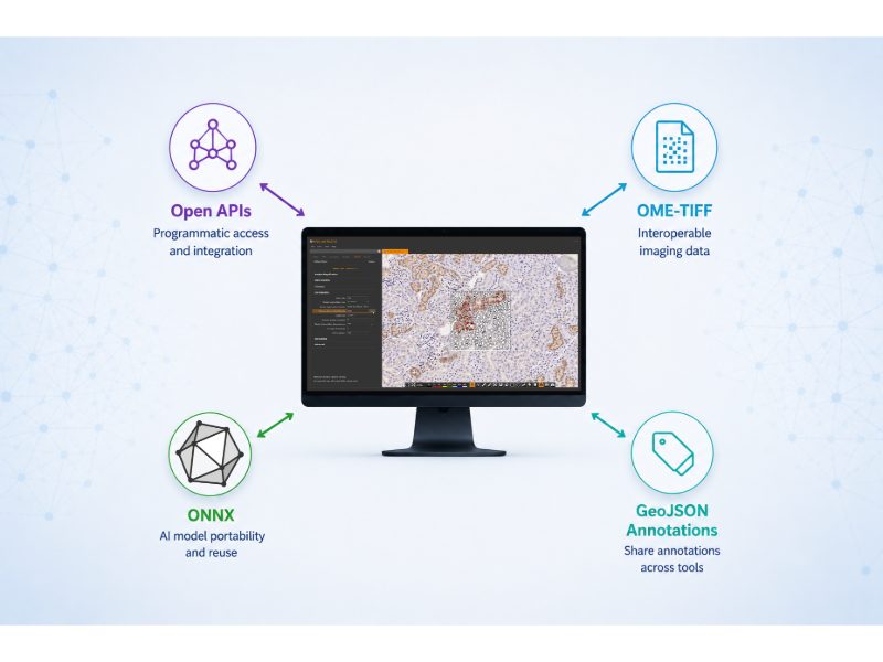
However, he acknowledges the field still has far to go, particularly regarding interoperability.
“We have a long way to go to achieve what pathologists and labs want — robust interoperability,” he said. “A lack of functional interoperability invites opportunity for medical error, which is what everybody wants to avoid.”
Looking Ahead
Dr. Wharton stated that while primary diagnosis is extremely important, image analysis algorithms and AI applied to the slides are truly the future.
“What we’re learning is that what AI recognizes in the slide image isn’t necessarily what the pathologist recognizes.”
Roche has taken steps toward this future through partnerships with over 12 algorithm companies in their Digital Pathology Open Environment.
“Looking to the future of precision digital diagnostics, Roche is in a great position to specify, evaluate, and provide the entire end-to-end in vitro diagnostic system workflow, from robust stain to WSI scan to viewing software to AI algorithm to results,” Dr. Wharton said. “This is what patients really need and what labs really want: confidence that when they assemble these complex systems from different components that they will give rise to an accurate and precise result for every patient.”
References
- Wharton KA Jr, Ranger-Moore J, Seng H, et al. Roche Digital Pathology Dx whole slide imaging system is comparable to traditional microscopy for primary diagnosis in surgical pathology. Am J Clin Pathol. Published online June 10, 2025. doi:10.1093/ajcp/aqaf052
No audio available for this article yet.
No quiz available for this article yet.