by Christos Evangelou, MSc, PhD – Medical Writer and Editor
Neural invasion occurs in 80%-100% of patients with pancreatic ductal adenocarcinoma (PDAC) and is an independent predictor of poor prognosis. However, the cellular mechanisms underlying neural invasion remain unknown. In a recent study, researchers at the Shenzhen Bay Lab, Peking University, Guangdong Provincial People’s Hospital, and Chongqing Medical University in China used cutting-edge single-cell and spatial transcriptomics to study the complex neural microenvironment in PDAC and identified cellular players that regulate neural invasion.
The study was published in Cancer Cell.
Studying the Neural Microenvironment of PDAC
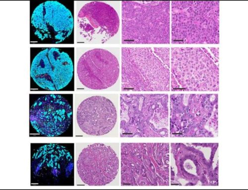
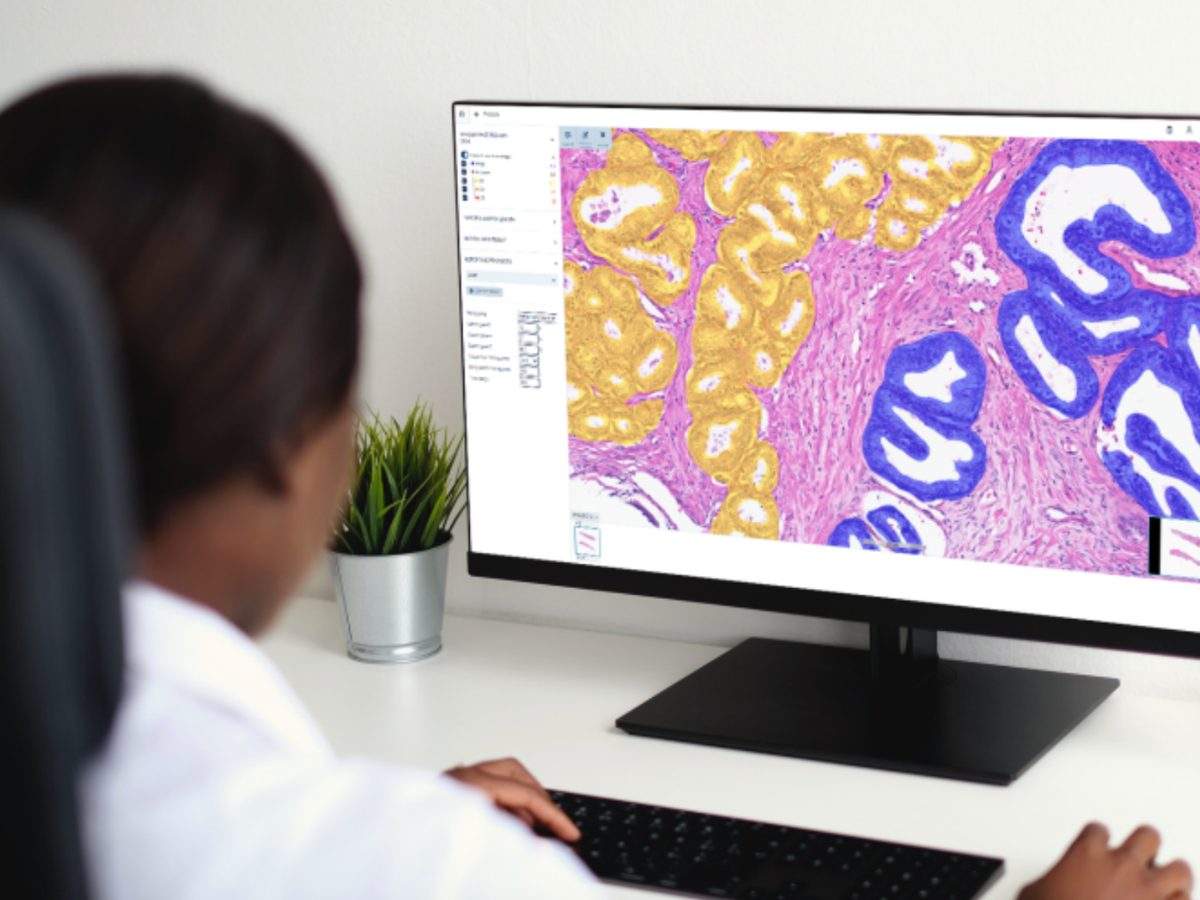
The research team integrated single-cell/single-nucleus RNA sequencing, spatial transcriptomics, and multiplex immunohistochemistry to analyze 62 samples from 25 treatment-naive patients with PDAC. This approach to creating a comprehensive map of tumor-associated nerves in PDAC enabled the team to study neural cells in tumor tissues in situ.
“Although pancreatic cancers are densely innervated, not all regions contain nerve bundles,” explained lead author Dr. Min-Min Chen of Shenzhen Bay Laboratory. “We performed H&E staining and pathological examination prior to region selection to ensure sufficient nerve-associated cells for analysis, and then used parallel tissue sections with multiplex immunohistochemistry to identify neuron populations while characterizing the complete neural ecosystem.”
The researchers classified samples into low neural invasion (8 samples) and high neural invasion (17 samples) groups, and analyzed differences in their cellular landscapes.
The Role of Tertiary Lymphoid Structures
One of the most notable differences between the low neural invasion and high neural invasion groups was the abundance and positioning of tertiary lymphoid structures (TLSs). In low neural invasion tissues, TLSs were abundant and positioned around non-invaded nerves, creating what the researchers term “nerve-TLSs.”
Spatial analysis revealed that approximately 50% of all TLSs detected in tumors were accompanied by nerve fibers within 100 micrometers. Among 141 non-invaded nerve fibers evaluated across nine tumor samples, 90 were associated with TLSs, while this proportion was low in invaded nerves.
“The interaction between nerves and TLSs presents a therapeutic opportunity,” noted Dr. Chen. “Strengthening the formation and function of TLSs in low neural invasion regions or promoting their establishment in high neural invasion patients might help boost local immune responses.”
According to Dr. Chen, the mechanism behind these differences appears to involve nerve-associated fibroblasts expressing high levels of CXCL12, which recruits CXCR4+ B cells and facilitates organization of TLSs. These structures support the expansion of CD8+ effector memory T cells (CD8_Temra-CX3CR1), which showed clonal expansion and migration from peripheral blood in patients with low neural invasion.
Promoters of Inflammatory Invasion
Tissues with high neural invasion exhibited a proinflammatory microenvironment dominated by NLRP3+ tumor-associated macrophages and cancer-associated myofibroblasts (myCAFs). These cells surrounded invaded nerves and created an environment characterized by T cell exhaustion rather than effective immune responses.
The researchers identified different macrophage populations within the nerves. Non-invaded nerves contained predominantly LYVE1+ tissue-resident macrophages, whereas invaded nerves showed high numbers of SPP1+ macrophages. This finding may suggest that resident macrophages change phenotype in response to cancer invasion.
TGFBI+ Schwann Cells
The research team identified three Schwann cell subsets and found that TGFBI+ Schwann cells were key promoters of neural invasion. Unlike the myelinating ABCA8+ Schwann cells or repair-associated SERPINA3+ Schwann cells, TGFBI+ Schwann cells localized at the leading edge of neural invasion and directly contacted invading cancer cells.
“Unlike repair Schwann cells, which were reported to be activated by c-Jun, TGFBI+ Schwann cells were induced by TGF-β from inflammatory macrophages and myofibroblasts,” Dr. Chen explained. “This suggests divergent differentiation pathways in regulating Schwann cell plasticity.”
Functional experiments demonstrated that TGF-β1 treatment of Schwann cells upregulated TGFBI and fibronectin expression while suppressing myelination markers. The resulting cells promoted pancreatic cancer cell migration and invasion in transwell assays, which could be blocked by TGF-β receptor inhibition. Survival analysis using external datasets demonstrated that TGFBI+ Schwann cell signatures correlated with poor prognosis in patients with PDAC.
Malignant Cell Heterogeneity and Invasion Potential
The study revealed that cancer cells within PDAC tumors had varying neural invasion potential. GABRP+ malignant cells showed the highest expression of nerve-related factors and were enriched near invaded nerves, while basal-like CEACAM6+ cells exhibited enhanced epithelial-mesenchymal transition signatures and integrin-mediated signaling pathways. A neuroendocrine-like APOA2+ subtype was exclusively found in tissues with low neural invasion and showed reduced expression of neural input signals, suggesting that certain malignant cell phenotypes may be inherently less invasive.
Clinical Implications and Future Directions
According to Dr. Chen, the identification of NLRP3+ macrophages as key inflammatory drivers suggests that targeting NLRP3 could inhibit neural invasion. She indicated that the TGF-β pathway is another potential therapeutic target, considering its role in transforming protective Schwann cells into invasion promoters.
“One potential strategy is to enhance the adaptive immune response in high neural invasion patients, possibly by targeting the factors contributing to macrophage-driven inflammation and T cell exhaustion,” Dr. Chen stated. “For the TGFBI+ Schwann cell pathway, our in vitro tests suggest that additional soluble mediators secreted by these cells may facilitate cancer cell migration synergistically.”
Dr. Chen emphasized that translating these findings into the clinic requires a move “from correlation to causation.” The team’s next steps include validation of findings in larger cohorts, functional analyses using patient-derived organoids or relevant animal models, and validation of biomarkers for clinical use.
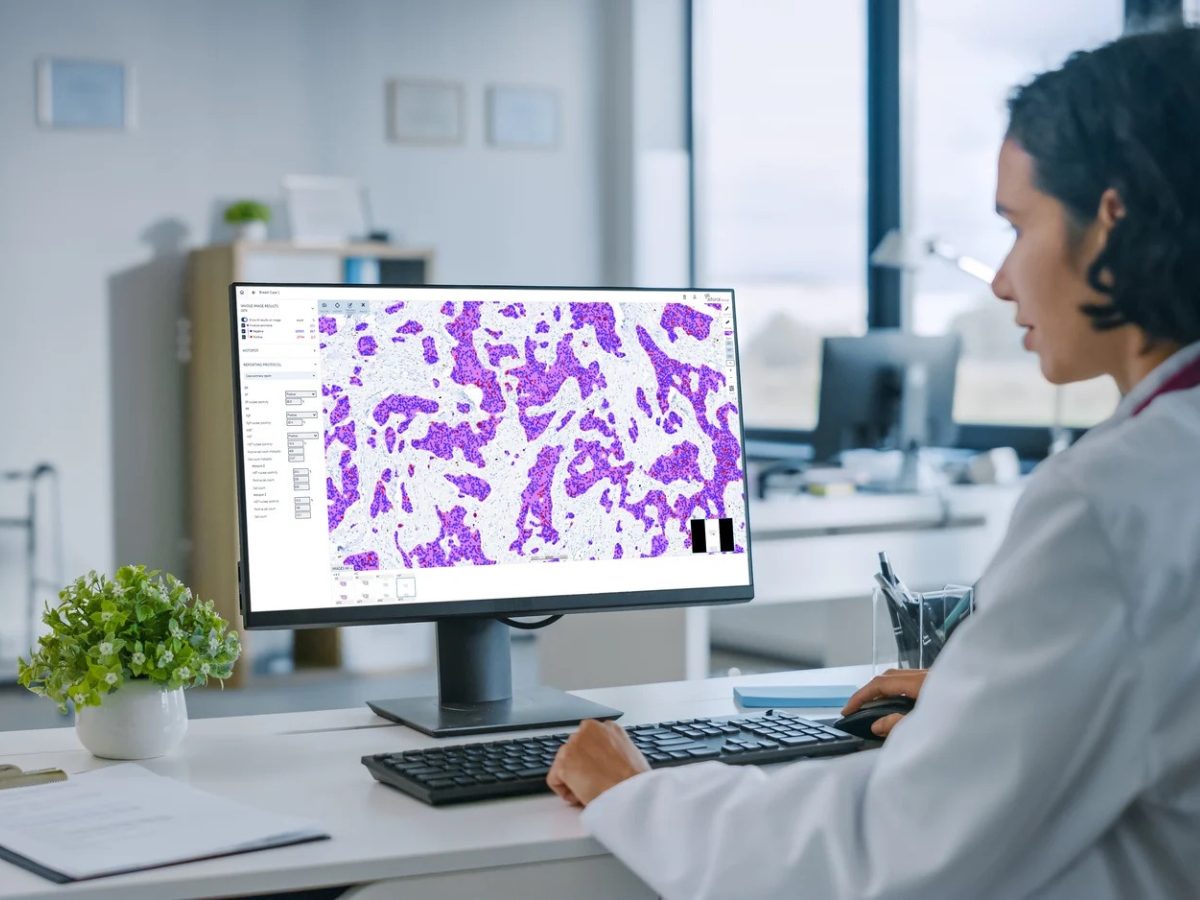
“Ideally, combining digital pathology with a panel of biomarkers could enable the identification of specific ‘bio-types’ of malignant cells, including those with high neural invasion potential,” Dr. Chen noted.
The study received financial support from the Major Program of Shenzhen Bay Laboratory, the National Key Research and Development Program of China, the National Natural Science Foundation of China, and the Open Program of Shenzhen Bay Laboratory.
References
- Chen MM, Gao Q, Ning H, et al. Integrated single-celland spatial transcriptomics uncover distinct cellular subtypes involved in neural invasion in pancreatic cancer. Cancer Cell. Published online July 17, 2025. doi:10.1016/j.ccell.2025.06.020
No audio available for this article yet.
No quiz available for this article yet.