by Christos Evangelou, MSc, PhD – Medical Writer and Editor
The Melanoma MEGA-Study, a decade-long study encompassing 1,653 patients from Hungary, integrated proteogenomics, AI-driven digital pathology, and clinical data to improve risk stratification for patients with melanoma. In an interview with Pathology News, Jeovanis Gil, PhD, one of the lead investigators of the Melanoma MEGA-Study, discussed how integrating proteogenomics, digital pathology, and AI-powered analytics can enhance our understanding of melanoma progression and therapy resistance.
Study Rationale
Approximately 10% of patients initially diagnosed with early-stage melanoma eventually progress to advanced disease; however, current prognostic tools fail to identify these high-risk patients. Gil and his international team found that traditional clinical factors, including the gold-standard Breslow thickness measurement, could predict only about half of recurrence cases.
“We saw that even thin, early-stage melanomas can recur unexpectedly, and current clinical tools like Breslow thickness simply don’t capture all the risk,” explained Gil. “This really highlights the limitations of relying just on pathology and patient history.”
This clinical gap became the driving force behind their initiative that would integrate proteogenomics, artificial intelligence, and spatial analysis to improve risk assessment in melanoma. “By layering in AI-driven spatial proteomics, we identified distinct molecular signatures, such as upregulation of mitochondrial protein translation and oxidative phosphorylation, that were much stronger predictors of recurrence.” The team previously published these findings in Clinical and Translational Medicine.
The research team analyzed samples from 1,653 patients with melanoma diagnosed between 2013 and 2022, with 361 patients contributing 819 metastatic tumor samples. This population-based cohort captured the entire melanoma burden from a single geographic region, providing insights into disease progression across all stages.
The Role of Mitochondrial Dysfunction in Melanoma Progression
Gil explained that one of the most striking results from the MEGA-Study is the central role of mitochondrial dysfunction in melanoma aggression, a finding that challenges traditional approaches focusing on genetic mutations alone.
“Mitochondrial pathways have emerged as a unifying theme,” Gil noted. “Across our studies, aggressive melanomas consistently upregulate mitochondrial protein synthesis and oxidative metabolism.”
In a study published in 2024, the team showed that BRAF-mutant metastatic tumors had elevated TCA-cycle and oxidative phosphorylation (OXPHOS) activity compared to wild-type tumors.
This work culminated in a 2025 study published in Cancer that demonstrated how the most proliferative melanomas “hyper-activate” the mitoribosome and OXPHOS pathways, creating a potential therapeutic vulnerability. The researchers found that blocking mitochondrial protein synthesis with certain antibiotics, originally designed to target bacterial machinery that closely resembles mitochondrial machinery, killed melanoma cells in laboratory studies.
“This particular work has got wide coverage worldwide, underscoring its impact,” Gil stated. “In short, these efforts spotlight mitochondria as an Achilles’ heel in melanoma.”
AI Meets Pathology
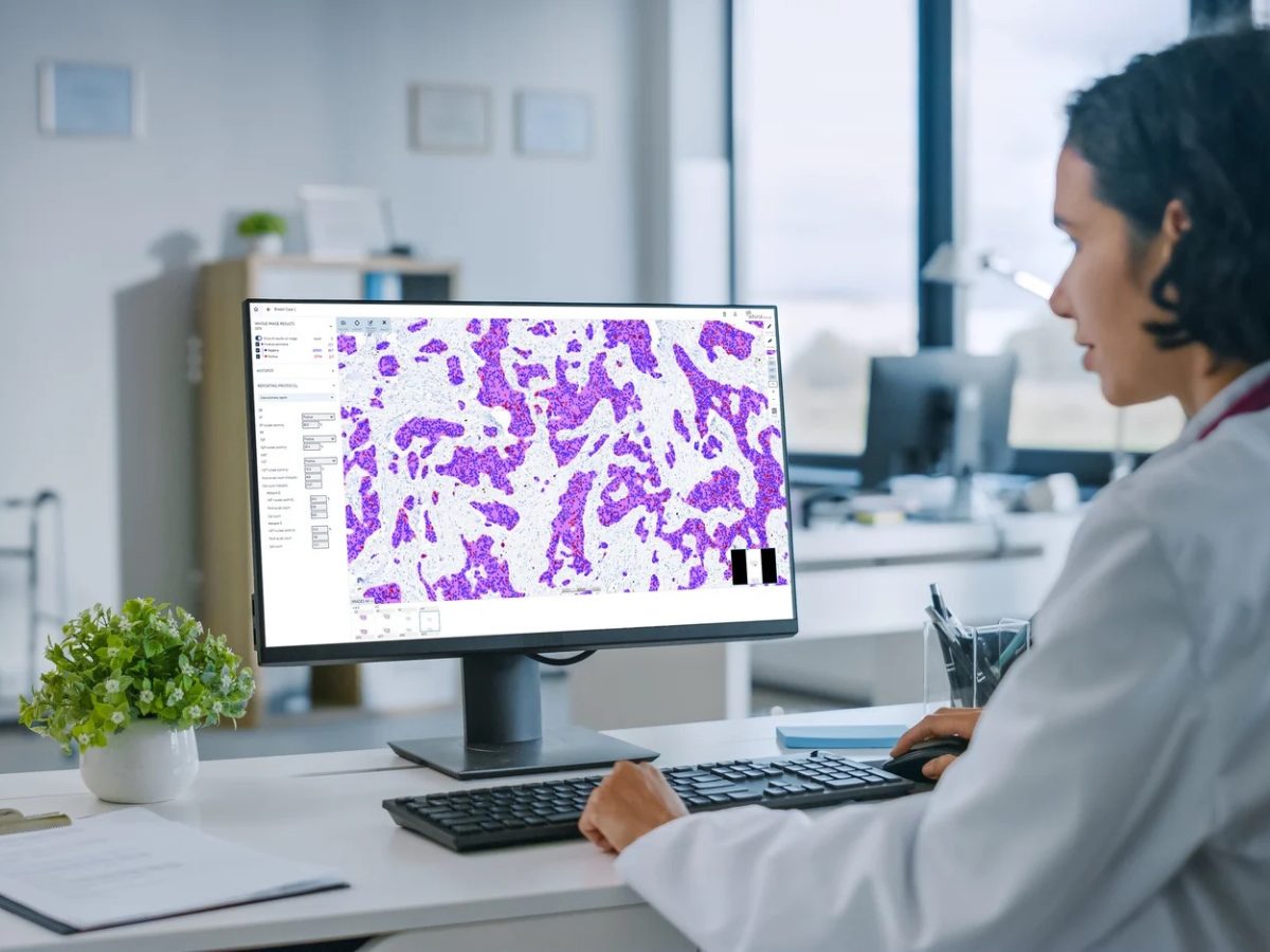
The research team harmonized various data streams, including proteogenomics, AI-driven digital pathology, and spatial proteomics. Specifically, the team developed an AI-powered digital pathology pipeline using the BIAS framework, training it to recognize tumor tissue, stromal regions, and normal skin with approximately 80% accuracy.
“Pathologists confirmed the AI was reliably distinguishing tumor cells from surrounding stroma and benign tissue,” Gil explained. “Building on that, we are now developing more advanced machine-learning models to go further; we’re using tools like QuPath and BIAS to mine image features across the whole cohort.”
The current models are being trained to predict recurrence risk or therapy response directly from histological patterns, integrating molecular and clinical data.
“After mastering ‘seeing’ the tumor architecture, we’re teaching the AI to read that architecture as a predictor of outcome,” he added.
This approach enabled the team to perform laser capture microdissection on specific tumor regions, isolating distinct compartments for downstream analysis. The resulting spatial proteomics revealed that recurrent melanomas show upregulated mitochondrial pathways in both tumor and stromal regions, whereas non-recurrent tumors exhibited immune-enriched microenvironments.
Proteomic Signatures Are Associated With Treatment Response
The MEGA-Study has identified proteomic signatures that can stratify patients into responders and non-responders for both targeted therapies and immunotherapy. Responders to immunotherapy displayed increased expression of proteins involved in antigen processing and immune system activation. In contrast, non-responders showed enrichment in DNA repair and RNA splicing pathways.
However, Gil is cautious about the clinical translation of these proteomic signatures.
“These biomarkers are not yet ready for clinical use; they remain in the discovery and early validation phase,” he acknowledged. “What’s promising is that our models consistently highlight the central role of mitochondrial activity in resistance, suggesting new avenues for combination therapies.”
He further explained that the pathway to clinical actionability involves a three-stage validation pipeline: confirming signatures in independent cohorts using targeted mass spectrometry and immunohistochemistry, testing biological relevance in preclinical models, and evaluating predictive power across diverse external populations.
Global Validation and Diversity
Although the single-center design of the MEGA-Study enabled consistent data collection over a decade, questions remain about generalizability across diverse populations. Gil noted that the research team is actively validating their findings in independent cohorts, both within Europe and globally.
The team is also collaborating with institutions in Mexico, analyzing samples from over 1,000 patients with melanoma, including many with acral and mucosal subtypes rare in European datasets, which, according to Gil, represents a crucial step toward addressing diversity in melanoma research.
“By including understudied groups, we can discover new molecular signatures specific to underrepresented melanomas, helping to close the gap in outcomes for patients who have historically been left out of research,” he explained.
Looking Ahead
As Gil noted,
“We urgently need in-depth molecular profiling to more accurately predict which early melanomas will relapse, and to help guide more personalized follow-up and treatment strategies,”
adding that the Melanoma MEGA-Study represents the possibility of more accurate risk stratification, better treatment selection, and ultimately, improved survival outcomes.
Gil revealed that, as part of the NIH’s Cancer Moonshot initiative and the International Cancer Proteogenome Consortium, the MEGA-Study’s approach is being extended to other aggressive cancers, including lung, gastric, and pancreatic tumors. Gil envisions a future where AI-driven digital pathology, in-depth molecular profiling, and advanced data analytics generate high-resolution molecular maps at the single-cell and microenvironment level across cancer types.
“For the field of precision oncology, real success means moving from ‘one-size-fits-all’ treatments to individualized strategies based on each patient’s unique tumor biology, regardless of cancer type or geographic location,” Gil stated. “Our hope, through these global collaborations, is to make true precision medicine accessible to all patients, not just in melanoma, but across the entire cancer spectrum.”
The MEGA-Study datasets have been made publicly available through the ProteomeXchange consortium and an interactive web interface at mmplot.com/melanoma.
The study received financial support from the Mrs. Berta Kamprad Foundation and the Crafoord Foundation in Sweden, and it was done under the auspices of a Memorandum of Understanding between the European Cancer Moonshot Lund Center and the U.S. National Cancer Institute’s International Cancer Proteogenome Consortium.
No audio available for this article yet.
No quiz available for this article yet.