by Christos Evangelou, MSc, PhD – Medical Writer and Editor
Foundation models trained on large datasets often produce outputs that require substantial refinement before they can be used reliably in clinical settings. To address this gap, researchers at Binghamton University developed ClinSegAI, a post-processing framework that refines outputs by correcting segmentation boundaries and producing masks suitable for downstream analysis without requiring retraining or fine-tuning of the underlying model. Validation experiments showed that ClinSegAI can improve the accuracy of cell segmentation in histopathology images.
The study was published in Computers in Biology and Medicine.
Study Rationale
Corresponding author Nancy Lan Guo, PhD, explained that pathologists often express the need for AI tools to streamline cancer diagnosis, particularly for identifying subtle patterns in medical images that are difficult for the human eye to detect.
“To address this need, we are leveraging the foundation model BiomedParse, which was trained on a large set of medical images that are otherwise inaccessible,” she said.
The researchers interviewed 40 pathologists from different academic and hospital settings to understand workflow priorities. Pathologists expressed the need for rapid identification of regions of interest on whole-slide images, slide-level triage to distinguish normal from tumor tissue, computation of radiomic features for prognostic modeling, and prediction of molecular features from histology images to make treatment decisions before sequencing results become available.
Methodology and Validation
The researchers evaluated ClinSegAI on multiple datasets containing images of various sizes and disease types. For primary testing, they used 128×128-pixel patches extracted from whole-slide images in The Cancer Genome Atlas (TCGA) for lung squamous cell carcinoma and adenocarcinoma. Ground truth annotations were created under supervision by a pathologist. For additional validation, they used the Triple-Negative Breast Cancer dataset (50 H&E images at 40× magnification), MoNuSeg (37 multi-organ images with 22,000 annotated nuclei), and NuInsSeg (H&E-stained 512×512 patches from 31 human and mouse organs).
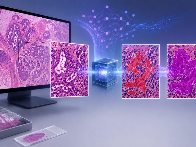
The pipeline operates in three stages. First, preprocessing is conducted to remove background noise through manual cropping in Photopea or semi-automated tissue isolation using Prov-Gigapath. Then, BiomedParse is used to generate initial probability maps for nucleus segmentation. Finally, ClinSegAI is used to apply proprietary refinement algorithms that correct boundaries, eliminate false positives, and separate merged cells or combine oversegmented regions.
Guo explained that, rather than building a segmentation model from scratch, the team enhanced BiomedParse through fine-tuning and a post-processing framework that incorporates additional information not included in the original training.
The researchers compared ClinSegAI against six widely used tools: Imaris (commercial microscopy software), CellViT (transformer-based approach), StarDist (star-convex polygon fitting), Fiji (open-source platform), Visiopharm (commercial digital pathology), and QuPath (open-source slide analysis).
“Metrics such as the Dice Similarity Coefficient (DSC), HD95, and ASSD were used to evaluate the accuracy of AI-based tumor boundary segmentation compared with ground truth, ensuring the quality of radiomics feature representation in downstream analyses,” Guo said.
Key Findings
ClinSegAI outperformed all other methods on the TCGA lung cancer dataset. The mean DSC was 0.79 for ClinSegAI, 0.69 for Imaris, 0.76 for Fiji, 0.59 for StarDist, and 0.61 for CellViT. Similarly, ClinSegAI outperformed other methods in most boundary alignment metrics on the TCGA lung cancer dataset.
External validation data suggest that the performance of ClinSegAI may be context dependent. On the TNBC and MoNuSeg datasets, StarDist achieved the best scores, which, according to Guo, was expected because these datasets were part of the training corpus of StarDist. ClinSegAI on these datasets demonstrated Dice Similarity Coefficients of approximately 0.79.
On the NuInsSeg dataset, which was not used to train any of the compared models, ClinSegAI demonstrated the highest DSC (approximately 0.79) and lowest boundary errors (HD95 around 30, ASSD of 6.5), with several pairwise differences reaching statistical significance. Fiji and StarDist showed mid-tier performance, while CellViT, QuPath, and Imaris showed larger errors.
For radiomic feature analysis, the team extracted more than 80 features including shape descriptors, first-order intensity measures, and texture calculations. Features from ClinSegAI-processed images showed fewer statistically significant differences from the ground truth than those from other methods. Methods producing merged cells yielded higher values for metrics such as sphericity or maximum intensity, whereas methods with boundary inaccuracies showed deviations in texture patterns.
Guo emphasized the multiple capabilities of the platform.
“The newly developed platform, ClinSegAI, enables comprehensive tumor analysis, combining segmentation, cell morphological assessment, and integration with spatial transcriptomics or other multi-omics data within a single platform,” she said.
Computational Considerations and Limitations
Runtime testing on a 1000×1000 MoNuSeg image showed approximately 3 seconds for segmentation and 27 seconds for radiomics computation across 275 cells (total 30 seconds), using 4GB GPU memory. Subsequent parallel processing reduced segmentation time to approximately 1 second.
Connected-component analysis, size filtering, and morphology operations add processing time and require more CPU/GPU memory. In routine use, total latency will depend on slide size, cell density, I/O bandwidth, and GPU availability. The team is pursuing vision-transformer-based parallel inference, allowing concurrent tile processing across multiple GPU streams while sharing a single transformer backbone, which should enable gigapixel whole-slide analysis within practical limits.
Future Work
Commenting on the pathway to clinical implementation, Guo said: “Evaluating the accuracy of ClinSegAI in both tumor cell classification and image segmentation against clinical standards and regulatory clearance are essential for its potential translation into real-world practice.”
She added that, by highlighting regions of interest on medical images, ClinSegAI can improve AI interpretability and build clinician trust. “As with other commercial software, ClinSegAI is designed to serve as an assistant, helping pathologists enhance workflow efficiency and diagnostic precision,” Guo said.
The study received financial support from the SUNY Empire Innovation Program and NSF 2444759.
References
- Bhajaj P, Nalubolu S, Gurram B, et al. ClinSegAI: A post-processing framework for superior histopathology segmentation accuracy, radiomics feature preservation, and quantitative analysis. Comput Biol Med. Published online November 11, 2025. doi:10.1016/j.compbiomed.2025.111298
No audio available for this article yet.
No quiz available for this article yet.