by Christos Evangelou, MSc, PhD – Medical Writer and Editor
A recent multi-omics study identified a previously unrecognized subtype of IDH-mutant astrocytoma that challenges traditional views of the role of immune infiltration in brain tumors. Despite showing extensive immune cell infiltration, typically associated with better outcomes, this newly defined subtype of immune/mesenchymal-enriched (IME) astrocytoma comprises approximately 13% of IDH-mutant astrocytomas and demonstrates worse prognosis.
The study was published in Cancer Cell.
Study Rationale
IDH-mutant astrocytomas are challenging brain tumor subtypes with high recurrence rates. Corresponding author Jiguang Wang, PhD, of Hong Kong University of Science and Technology, explained that genomic and transcriptomic analyses do not fully capture the biological complexity of gliomas.
“Genomic and transcriptomic frameworks capture mutations and RNA expression signals, but many oncogenic programs and therapeutic targets are executed at the protein level,” Wang noted. “We repeatedly observed poor mRNA–protein concordance for key pathways involving immune signaling, extracellular matrix remodeling, and metabolic rewiring.”
This knowledge gap prompted the research team to conduct a proteogenomic analysis of IDH-mutant astrocytomas, integrating bulk multi-omics data from 51 patients with single-cell RNA sequencing, spatial transcriptomics, and advanced imaging techniques.
Molecular Subtypes
The researchers used unsupervised clustering of proteomics data and identified four molecular subtypes of astrocytomas: adipogenesis/fatty acid metabolism (AFM), proliferative/progenitor (PPR), immune/mesenchymal-enriched (IME), and neuronal (NEU). Each subtype demonstrated distinct biological characteristics and clinical outcomes.
The AFM subtype showed enrichment for metabolic programs such as lipid and fatty acid metabolism. PPR tumors were characterized by cell cycle activation and DNA replication pathways, with a high prevalence of CDKN2A/B deletions, which are associated with poor prognosis. The NEU subtype exhibited enhanced neuronal functions, including synaptic transmission. The IME subtype showed enrichment in interferon-γ response signatures and epithelial-mesenchymal transition programs.
To validate these protein-based classifications across larger cohorts lacking proteomics data, the team developed a transcriptomics-based classifier. When applied to independent cohorts from The Cancer Genome Atlas (TCGA, n=234) and the Chinese Glioma Genome Atlas (CGGA, n=273), the classifier achieved high accuracy in distinguishing the four subtypes.
The Paradox of Immune-Hot but Aggressive Tumors
PPR tumors showed poor outcomes, which, according to Wang, was expected because of their association with grade 4 features and CDKN2A/B deletions. IME tumors also demonstrated aggressive behavior despite lacking these traditional high-risk markers. In multivariable Cox models adjusting for grade, age, and gender, IME remained an independent predictor of poor prognosis across multiple cohorts.
Single-cell RNA sequencing analysis provided insights into potential biological mechanisms behind the paradoxically poor prognosis of IME tumors. IME tumors were enriched in lymphocytes, especially exhausted T cells and plasma cells.
“Our observation showed that having more immune cells equals better outcomes does not hold universally,” Wang explained. “Prognosis depends on quality and context such as T-cell exhaustion, suppressive myeloid programs, and ECM barriers, rather than sheer infiltration.”
The team went one step further to analyze the spatial organization of immune cells infiltrating IME tumors using 10x Visium HD spatial transcriptomics and CODEX spatial proteomics. They found that IME tumors contained distinct perivascular lymphocytic cuffing structures, where exhausted T cells and plasma cells clustered around the blood vessels.
Gemistocytic Differentiation as a Diagnostic Marker
The researchers analyzed the histopathological features of the different molecular subtypes of astrocytomas. They found that IME tumors showed gemistocytic differentiation, characterized by tumor cells with enlarged, eosinophilic cytoplasm and eccentric nuclei. This morphological feature was present in all IME cases in the discovery cohort and was validated using large language model-assisted analysis of pathology reports from TCGA cases. Wang explained that these features resemble reactive astrocyte states. “We hypothesize these tumors mirror a reactive astrocytoma,” he noted.
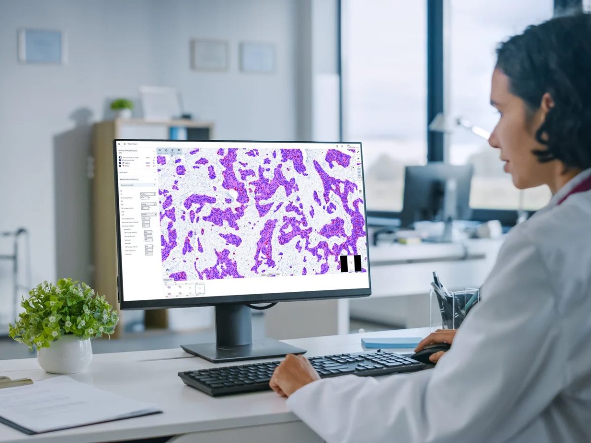
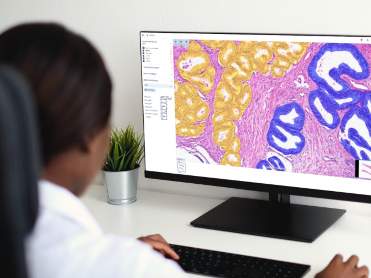
The researchers developed an AI model capable of identifying gemistocytic differentiation from routine histopathology images. The AI classifier strongly correlated with pathologist gemistocyte quantification (Pearson correlation coefficient = 0.970) and achieved high performance in classifying IME (area under the curve = 0.927).
“This provides a practical surrogate marker for IME tumors, particularly in settings without access to multi-omics profiling,” Wang stated.
Evolutionary Dynamics and Tumor Progression
Longitudinal analysis of 189 initial-recurrent tumor pairs showed that the frequency of the AFM subtype decreased at recurrence. In contrast, both PPR and IME subtypes became more prevalent at recurrence, suggesting that they represent more aggressive evolutionary trajectories. In addition, although gemistocytic differentiation was conserved during tumor evolution, recurrent IME tumors showed increased fibrous hyperplasia and fibroblast infiltration, indicating continued microenvironmental remodeling.
Mechanistic Insights Point to Therapeutic Targets
The research team assessed the key molecular drivers of IME aggressiveness and found that interferon-stimulated genes, particularly GBP1 and GBP2, were upregulated in IME tumors. Functional experiments demonstrated that GBP1 overexpression increased proliferation and migration in IDH-mutant astrocytoma cell lines, suggesting direct tumor-promoting effects. The team also found elevated expression of immunoglobulin components and plasma cell markers in IME tumors, with spatial analysis confirming colocalization between plasma cells and tumor cells.
GUIDE: An AI-Powered Diagnostic Platform
To facilitate clinical implementation, the researchers developed GUIDE (Generic Utility for IME Diagnostic Estimation), an AI-powered multi-omics diagnostic platform that can integrate any available data types.
“GUIDE addresses the reality of clinical settings where missing modalities are common,” Wang explained. “Rather than imputing missing values, GUIDE is trained directly on datasets with missing modalities and generates predictions based on available data.”
The platform achieved high performance across different data availability scenarios, with imaging alone providing ~84% balanced accuracy, improving further when combined with molecular data.
Potential Clinical Implications
Wang emphasized that
“checkpoint blockade alone is unlikely to suffice in patients with the IME subtype” because these tumors have profound T-cell exhaustion and an immunosuppressive tumor microenvironment. He added that combination approaches are more likely to succeed, explaining that “PD-1/PD-L1 paired with myeloid reprogramming or tumor-targeted strategies and dual TGF-β/VEGF blockade are needed to normalize stroma and vasculature and improve T-cell trafficking.”
Wang also suggested that direct plasma cell targeting using anti-CD38 or BCMA-directed approaches might be beneficial, although this approach remains speculative given potential risks to systemic immunity and uncertain CNS penetration.
Limitations and Next Steps
The authors acknowledged that the discovery cohort was relatively small (n=51), although validation in larger independent cohorts strengthened the findings. They also acknowledged differences in ancestry and treatment between the CGGA and TCGA cohorts, which may influence glioma biology. Moreover, the spatial analyses focused on tumor cores rather than invasive margins, which may be important for understanding tumor progression.
Wang commented on their plans for expanding GUIDE:
“We will extend GUIDE to additional glioma subtypes and build multi-institutional cohorts with prospective sample collection to confirm subtype prevalence and prognostic value.”
The team is also preparing biomarker-driven clinical trials targeting the IME subtype.
As Wang concluded by saying,
“Our goal is pragmatic: to deliver a classification that not only explains biology but also guides real-world decisions with assays feasible in routine clinics.”
The open-science approach, with model weights and protocols being made freely available, ensures that these advances can benefit patients worldwide.
The study received financial support from the Noncommunicable Chronic Diseases-National Science and Technology Major Project, NSFC/RGC Collaborative Research Scheme, National Natural Science Foundation of China, RGC grants, ITC grants, CASCroucher Funding Scheme, and the Beijing Municipal Health Commission.
References
- Tang J, Fan W, Ruan Y, et al. Protein-based classification reveals an immune-hot subtype in IDH mutant astrocytoma with worse prognosis. Cancer Cell. Published online September 11, 2025. doi:10.1016/j.ccell.2025.08.006
No audio available for this article yet.
No quiz available for this article yet.