by Christos Evangelou, MSc, PhD – Medical Writer and Editor
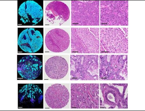
A new computational approach developed by researchers at Karolinska Institutet improves the resolution of breast cancer spatial analysis by integrating AI-powered image analysis with spatial transcriptomics. The pipeline, which the researchers termed “computational tissue annotation” or CTA, uses machine learning to identify individual cell types in standard H&E-stained tissue sections, providing spatial insights into cellular neighborhoods and their molecular signatures in breast tumors.
The study was published in NPJ Precision Oncology.
Study Rationale
Spatial transcriptomics analysis helps researchers understand how genes are expressed within tissues. However, first author Dr. Tianyi Li of Karolinska Institutet explained that current technologies face resolution challenges. Visium, a widely used spatial transcriptomics platform, captures gene expression data from spots 55 micrometers in diameter, which is large enough to encompass multiple cells of different types, creating mixed gene expression profiles that do not reveal individual cellular contributions.
“The platform’s relatively large spot size often encompasses multiple cells, generating a mixed gene expression profile,” Li noted. “This spot-based approach limits the ability to resolve the contribution of individual cells to the tumor microenvironment.”
The research team recognized that the rich morphological information present in routine H&E images could be leveraged to increase the spatial resolution of current spot-based spatial transcriptomics platforms.
Combining Pathology Expertise with Machine Learning
The research team combined the QuPath platform, an open-source tool widely used in research and clinical pathology, with a random trees algorithm for cell classification. Li explained that this approach was chosen for its accessibility and performance characteristics.
“We choose the random trees algorithm for cell classification because it performs well with the small training sets and leverages a wide range of features during the model training,” Li stated.
The algorithm incorporates multiple histological features including nuclear area, cell area, circularity, and staining optical density to construct robust cell type predictions.
The researchers validated the CTA pipeline using 23 breast tumor sections from four patients, two of whom had triple-negative or HER2-positive tumors. All computational annotations were independently reviewed by board-certified pathologists to ensure clinical reliability.
Performance of Deconvolution Methods
The researchers conducted an evaluation of widely applied deconvolution methods, which are computational approaches that attempt to separate mixed cell type signals in spatial data. Using CTA results as a reference, the team compared seven commonly used deconvolution methods across tumor, immune, and stromal cell populations.
Cell2location, RCTD, and Stereoscope showed the strongest correlations with morphology-based annotations, with median correlation coefficients exceeding 0.65 for tumor cells. According to Li, these findings provide clinicians and researchers with evidence-based guidance for selecting appropriate analytical tools for their spatial transcriptomics studies.
CTA Provides Enhanced Spatial Resolution
The CTA pipeline provided enhanced resolution in tumor analysis, revealing previously unknown aspects of breast cancer biology. Even clusters that appeared homogeneous based on gene expression analysis contained substantial cellular heterogeneity when examined using the CTA method.
For example, in one HER2-positive sample, a cluster dominated by immunoglobulin-encoding genes, which are typically associated with B cell populations, contained substantial levels of stromal cell populations that would have been missed using traditional spot-based analysis methods. According to Li, this finding demonstrates how relying solely on top-expressed genes can sometimes lead to biased cluster annotations.
The CTA pipeline also enabled high-resolution copy number variation (CNV) analysis by using quantitative tumor purity cutoffs.
“By incorporating these cutoffs, the CTA pipeline can help to minimize the likelihood of cancer cell contamination in germline spot selection and refine the CNV calling by increasing the tumor purity threshold,” Li explained.
In one triple-negative breast cancer sample, this approach identified distinct clonal spatial distributions using a 90% tumor purity cutoff.
Potential Implications
According to the authors, the CTA pipeline can help researchers identify spatially resolved breast cancer subtypes and uncover patterns of intrinsic subtypes within individual tumors, which may be missed by standard spatial transcriptomics methods. In addition, the pipeline enhanced visualization of lymphocyte clone interactions within the tumor microenvironment.
“By integrating our CTA pipeline with Spatial VDJ technology, we identify the tumor-immune interaction as well as B cell and T cell clones localized within the tumor niches,” Li said. “The enhanced resolution in the visualization allows the detection of potential B cell clones that may serve as candidates for further development of immune therapeutics.”
Limitations and Future Work
The researchers acknowledge that the CTA pipeline requires high-quality original images and can be influenced by tissue quality, staining protocols, and imaging conditions. In addition, the study focused exclusively on ductal carcinomas, and the performance of the CTA method in lobular cancers or rarer breast cancer subtypes remains unknown.
Technical challenges also exist, particularly with fresh-frozen sections used in spatial transcriptomics, which often have thicker sections and more ambiguous morphology compared to standard diagnostic slides. However, fresh-frozen tissues preserve RNA quality better, which is important for spatial transcriptomics analysis.
The research team is currently expanding the capabilities of the CTA pipeline to newer platforms.
“We are working on implementing the CTA pipeline on the VisiumHD and Xenium platforms,” Li revealed.
These newer spatial technologies offer even higher resolution, potentially amplifying the benefits of morphology-assisted analysis.
The researchers are also working to expand the applications of the CTA method to other cancer types.
“We believe that our CTA outputs can also complement the gene expression analysis by incorporating the histopathological information into the data analysis pipeline,” Li concluded.
The study received financial support from Karolinska Institutet.
References
- Li T, Yang Q, Acs B, et al. Computational pathology annotation enhances the resolution and interpretation of breast cancer spatial transcriptomics data. NPJ Precis Oncol. 2025;9(1):310. Published 2025 Sep 9. doi:10.1038/s41698-025-01104-3
No audio available for this article yet.
No quiz available for this article yet.