by Christos Evangelou, MSc, PhD – Medical Writer and Editor
In a recent pan-cancer study, researchers from Chongqing Medical University, the Changping Laboratory, and Peking University used integrated spatial transcriptomic profiling to characterize the cells that constitute tumor-associated tertiary lymphoid structures. The study revealed not just what cells are present in these immune structures, but where they’re located and how they interact to create functional antitumor responses, pinpointing pathways that could transform immunologically ‘cold’ tumors into treatment-responsive ‘hot’ ones.
In an interview with Pathology News, corresponding author Sijin Cheng, PhD, of Chongqing Medical University, talked about the rationale behind their study, the technical challenges they faced, and the implications of their findings.
The study was published in Cell Reports.
Study Rationale
Some tumors develop organized clusters of immune cells called tumor-associated tertiary lymphoid structures (TA-TLSs), which can initiate antitumor immune responses. These structures resemble miniature lymph nodes and have been associated with improved patient outcomes and stronger responses to immunotherapy.
“Tumor-associated tertiary lymphoid structures play a crucial role in regulating antitumor immune responses and influencing the efficacy of immunotherapy,” Cheng explained. “However, our understanding of how TA-TLSs form remains limited, particularly regarding the key cell types that drive their induction.”
Methodology
The research team integrated spatial transcriptomic data from 517 samples across 23 different cancer types to create a pan-cancer atlas of TA-TLSs. The inclusion of samples from multiple cancer types allowed them to identify shared characteristics of TA-TLS while also revealing cancer-type-specific variations.
CCL19+ Perivascular Cells as Potential Lymphoid Tissue Organizer
The researchers identified a previously underappreciated cell type: CCL19+ perivascular cells. Cheng explained that these cells tend to wrap around blood vessels within tumors and appear to function as lymphoid tissue organizer cells, recruiting and coordinating immune cells to build TA-TLSs.
“From a clinical translation perspective, the discovery of CCL19+ perivascular cells is particularly paradigm-shifting,” Cheng stated. “Their innate perivascular location positions them ideally for lymphocyte recruitment. Existing research suggests that perivascular cells possess significant plasticity and can differentiate into key reticular stromal cells that provide the structural foundation for TA-TLSs.”
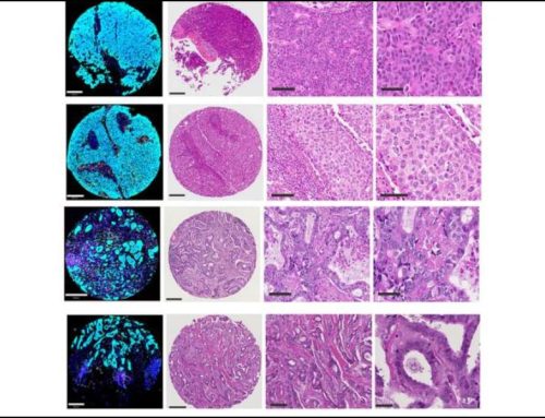
The research team validated their computational findings through multiple experimental approaches, including immunofluorescence staining, which confirmed that these CCL19+ cells cluster near lymphocyte-rich regions. Areas with CCL19+ perivascular cells showed significantly more B cell infiltration compared to regions lacking CCL19+ perivascular cells, supporting their role as immune recruiters.
Arterial Transformation
The research team found that arterial endothelial cells within TA-TLSs can undergo transformation, acquiring characteristics similar to those of high endothelial venules (HEVs), which are specialized blood vessels that facilitate the entry of immune cells into lymphoid organs.
The team used trajectory analysis of spatial transcriptomic data to track how arterial endothelial cells gradually lose their arterial identity and acquire HEV-like properties, including the enhanced expression of adhesion molecules that facilitate the attachment and transmigration of immune cells across blood vessel walls. Cheng explained that the transformation of arterial endothelial cells within TA-TLSs appears to be mediated by downregulation of NOTCH1 signaling, a finding the team validated through laboratory experiments.
“We observed arterial endothelial cells present within TA-TLSs, which could undergo a transformation to acquire HEV-like phenotypes,” Cheng said. “Our findings suggest that manipulating NOTCH signaling could potentially enhance immune cell recruitment into tumors.”
The Role of TA-TLS Maturation
The study also provided insights into how the maturation stage of TA-TLSs affects their antitumor function. The research team classified TA-TLSs into three maturation stages: lymphoid aggregations (LAs), primary follicle-like structures (PFLs), and secondary follicle-like structures (SFLs) containing germinal centers.
They found that mature TA-TLSs produced IgG antibodies rather than IgA antibodies. This antibody preference was consistent across multiple cancer types, which, according to Cheng, indicates a fundamental principle of TA-TLS function and suggests that mature TA-TLSs are optimized for systemic antitumor responses rather than mucosal immunity.
In addition, mature TA-TLSs showed clear compartmentalization, with B cells concentrated in central regions and plasma cells positioned at the periphery. This arrangement facilitates antibody production and dissemination into surrounding tumor tissue, Cheng explained.
Technical Innovation and Potential Clinical Implications
The study relied on advanced spatial transcriptomics, specifically the 10X Visium platform, which allows researchers to measure gene expression while preserving information about where cells are located within tissue samples.
“Integrating spatial transcriptomic data generated from different studies posed one of the most significant technical challenges,” Cheng acknowledged.
The team overcame this by optimizing computational methods and validating their findings through multiple experimental approaches, including parallel analysis of tissue sections using both spatial transcriptomics and traditional immunofluorescence staining.
“The primary challenge for treating ‘cold’ tumors is enhancing their immune infiltration,” Cheng stated. “Our study addresses this by characterizing TA-TLSs and identifying key stromal cells critical for their formation. This opens up a promising strategy: targeting these specific cell types to induce TA-TLS development, thereby converting immunologically ‘cold’ tumors into ‘hot’ ones.”
Cheng explained that a potential therapeutic approach would be to target perivascular cells to promote their differentiation into CCL19+ organizer cells, which could stimulate TA-TLS formation. Combining NOTCH pathway inhibitors with existing immunotherapies might enhance immune cell recruitment by promoting arterial-to-HEV transformation, he added.
Future Directions
Cheng revealed that the team plans to validate their findings in mouse models and test whether manipulating CCL19+ perivascular cells or arterial endothelial cells in vivo can indeed promote TA-TLS formation and improve tumor control.
“The critical next step is to assess their causal roles and functional impact on tumor progression using in vivo mouse models,” Cheng stated. “We can develop antibody-based strategies to specifically target perivascular cells, aiming to induce their differentiation into CCL19+ perivascular cells.”
The team also plans to explore combination therapies, specifically testing whether NOTCH pathway inhibitors can enhance the effectiveness of immune checkpoint blockade therapy in tumors that are resistant to treatment.
The study received financial support from the Start-up Grant for Recruited Scholars of Chongqing Medical University, the Chongqing Education Commission of China, the Changping Laboratory, the National Science and Technology Major Project, the National Key Research and Development Program of China, the CAMS Innovation Fund for Medical Sciences, and the Beijing Natural Science Foundation.
References
- Li X, Chu X, Xu W, et al. Integrated spatial transcriptomic profiling to dissect the cellular characteristics of tumor-associated tertiary lymphoid structures. Cell Rep. 2025;44(9):116250. doi:10.1016/j.celrep.2025.116250
No audio available for this article yet.
No quiz available for this article yet.