by Christos Evangelou, MSc, PhD – Medical Writer and Editor
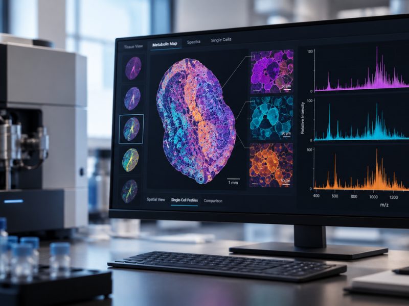
Researchers at the University of Münster and Bruker Daltonics GmbH & Co. KG in Germany developed a novel imaging technique that combines mass spectrometry with integrated microscopy to study cellular behavior at the molecular level. The method can map lipids and metabolites within individual cells in intact tissue, providing spatial metabolic insights at the single-cell level. According to the authors, this method can help researchers better understand disease processes by revealing metabolic signatures that conventional imaging methods cannot detect.
The study was published in Nature Communications.
Study Rationale
Jens Soltwisch, PhD, researcher at the University of Münster and the corresponding author of the study, explained that spatial analysis of small molecules, particularly lipids and metabolites, has lagged behind methods for analyzing spatial gene expression. However, genes and proteins tell only part of the story; lipids and metabolites are the downstream products that affect cellular metabolism and behavior.
“While transcriptomics and proteomics are already widely used in spatial biology, the analysis of small molecules like lipids and metabolites on the single-cell level is still in an early stage of development,” Soltwisch said in an interview with Pathology News. “In my opinion, spatially resolved analysis of these small molecules is needed because it delivers essential information for a more holistic understanding of biological processes.”
Soltwisch noted that technical challenges have contributed to this gap in spatial lipidomic analyses. Although matrix-assisted laser desorption/ionization mass spectrometry imaging (MALDI-MSI) can be used to detect metabolites, achieving single-cell resolution while maintaining co-registration with microscopy images is difficult. Previous efforts involving analysis on separate tissue sections were limited by alignment errors that made cellular-level analysis less accurate.
Methodology
The team developed a transmission-mode MALDI-2-MSI system with integrated bright-field and fluorescence microscopy built into the ion source. Soltwisch explained that this design allowed them to perform both microscopy and mass spectrometry imaging on the same tissue section using a single coordinate system, overcoming co-registration challenges.
“Working on the micron and sub-micron scale, precise positioning of the sample in all three dimensions becomes very important,” Soltwisch stated. “Luckily, we were able to work in a very tight and fruitful collaboration with Bruker Daltonics to construct and build the new ion source with the necessary precision.”
The system achieves pixel sizes of 1 × 1 micrometers, which are small enough to resolve subcellular structures. The researchers also developed staining protocols that enable immunofluorescence analysis before mass spectrometry imaging without compromising the chemical integrity of lipids and metabolites.
The integrated system uses in-source fluorescence microscopy as an intermediate step to link external high-resolution microscopy images with mass spectrometry data. This achieves co-registration accuracy of less than 1 micrometer, allowing for single-cell analysis.
System Validation
To validate their method, the researchers examined macrophages engulfing bacterial particles. By combining fluorescence markers that light up when bacteria are digested with mass spectrometry imaging, the team could map the chemical changes occurring inside individual phagolysosomes, the cellular compartments where pathogens are destroyed.
The analysis revealed that bacterial lipids are broken down into specific metabolites, including lysophosphatidylglycerols, which result from the enzymatic degradation of bacterial membrane components. According to Soltwisch, this level of detail — tracking metabolic processes within subcellular compartments — was previously unattainable.
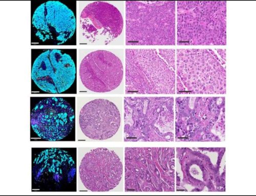
To further validate their system for a different application, the team used it to analyze tumors isolated from a mouse model of aggressive breast cancer. The researchers analyzed more than 63,000 individual cells in a single tissue section and identified eight distinct tissue microenvironments and eight subtypes of neutrophils based on their lipid profiles.
The researchers combined morphometric data with immunofluorescence markers for immune cells (CD45), neutrophils (Ly6G), and T3 neutrophils (DcTRAIL-R1). This allowed the team to track how neutrophil metabolism varies across different regions of the tumor. Some neutrophil subtypes showed lipid profiles similar to those of their surrounding tissue, suggesting active uptake and recycling of lipids from the microenvironment. Other subsets maintained metabolic signatures distinct from those in their surroundings.
“I was surprised by how much variation can be found in the lipid profile of neutrophils in cancer and how much it seems to interact and adapt with and to its direct microenvironment,” Soltwisch said.
In addition, the team found that some neutrophils clustered in hypoxic regions of the tumor. These cells showed elevated levels of acylcarnitine, a metabolic marker of hypoxia, as well as increased levels of saturated lysophosphatidylcholines, which have been implicated in metastasis and immune cell recruitment. The T3 neutrophil subset, which concentrates in hypoxic areas of the tumor, displayed a metabolic signature that was distinct from the metabolic profiles of other neutrophil subsets. The researchers also found that neutrophils near adipose tissue showed increased levels of triglycerides and diglycerides, whereas those deeper in the tumor did not.
Potential Applications
Soltwisch explained that mapping the metabolic heterogeneity of tumor-infiltrating immune cells could help explain why some tumors respond to immunotherapy while others do not. The lipid signatures identified across different neutrophil subtypes might serve as biomarkers of treatment response or as targets for metabolic intervention. The technique could also be used to study how immune cells adapt their metabolism during pathogen clearance.
“I think the technique has the potential to introduce a new ‘column’ or two into multiomics datasets,” Soltwisch noted. “In the best case, it can deliver the direct analysis of metabolic or lipid pathways and provide complementary information to gene regulation and protein expression.”
Future Directions
Despite the promising validation findings, the current sensitivity restricts analysis primarily to abundant lipid species; detecting low-abundance metabolites remains challenging. The method is also limited to molecules that ionize efficiently with MALDI.
“Our next steps will center around the application of the technique to a number of pressing analytical questions to see how much impact the technique can have to answer them,” Soltwisch stated. “We are also working on extending the technique towards the analysis of a broader range of metabolites. Of course, higher spatial resolution and higher sensitivity are always better, so we are also thinking about ways to improve in those fields, as well.”
The team plans to explore whether combining this approach with complementary techniques, such as plasma-based ionization, could expand the range of detectable metabolites. Increasing throughput will also be important for clinical applications, where analyzing multiple patient samples efficiently is essential.
References
- Potthoff A, Schwenzfeier J, Niehaus M, et al. Spatial biology using single-cell mass spectrometry imaging and integrated microscopy. Nat Commun. 2025;16(1):9129. Published 2025 Oct 15. doi:10.1038/s41467-025-64603-8