

Register using the link below
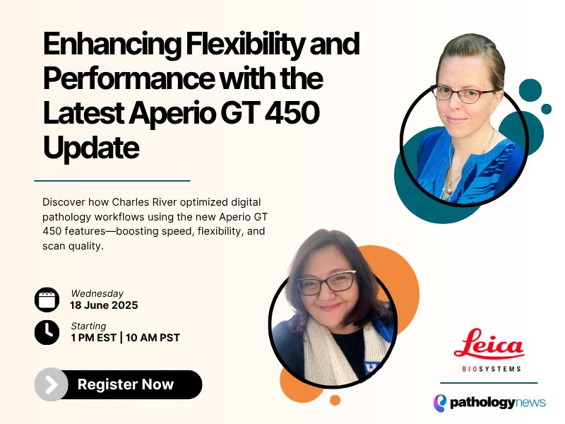
Recent updates to the Aperio GT 450 introduce exciting new capabilities, including scanning at 20x, z-stack acquisition, and manual scanning options for challenging slides. These enhancements bring even greater flexibility to the Aperio GT 450 platform, building on its well-established speed and throughput advantages.
Charles River Laboratories, a long-time user of the Aperio AT2 with over a dozen units in operation at peak use, had previously relied on manual scanning capabilities for certain complex tissue types such as bone marrow, ocular tissue, small samples, and cell lines. The new Aperio GT 450 enhancements offer a bridge between the flexibility of the Aperio AT2 and the high-speed automation of the Aperio GT 450, giving laboratories even more tools to meet diverse scanning needs.
As a beta site for the enhanced features, Charles River Laboratories evaluated the latest Aperio GT 450 functionality and found the manual scanning capabilities particularly impactful. Based on their positive experience, they are now moving forward with upgrading their entire Aperio AT2 fleet to the enhanced Aperio GT 450 platform.
In this presentation, we’ll deepen your understanding of the Aperio GT 450 and explore real-world examples of the updated manual scanning features compared with legacy Aperio AT2 scans. We’ll also share strategies for optimizing lab workflows for digital pathology, offer practical tips for scanning, especially with difficult tissue types, and discuss the broader value of going digital.
Learning Objectives:
- Deepen understanding of the Aperio GT 450 platform and its enhanced features
- Learn how to optimize your lab for scanning success—workflow, lab design & slide preparation
- Understand how slide prep quality impacts downstream imaging
- Explore tips and techniques for scanning difficult tissues
- Discuss the benefits and opportunities of digital pathology
For research use only. Not for use in diagnostic procedures.
Presenters
Sarah O’Connor
Sarah O’Connor has almost 10 years of experience in digital pathology and has utilized multiple scanning platforms. As a Digital Pathology Implementation and Training Specialist with Charles River Laboratories, Sarah has supports Charles River’s efforts to further their digital delivery through product testing and validation of new scanners and digital pathology software.
Charles River’s digital pathology process is fully validated for GLP primary evaluation and peer review use. During her tenure as a Tissue Cross Reactivity Research Assistant Sarah gained extensive experience with hard to scan tissue such as cell lines, protein spots, and faintly stained tissues. When not focused on digital pathology, Sarah likes to spend her time hiking and fly fishing with her dog Beowulf.
Diane Robirds
Diane Robirds joined Leica Biosystems in 2015 and has worked in Digital Pathology sales, training, and application support roles. Before joining Leica Biosystems, she spent many years in Genomics and Pathology laboratories, both clinical and research, from the bench to lab management.
In her current role as National Workflow and Solution Architect for Life Sciences, she enjoys getting to know each laboratory’s unique goals and needs on their Digital Pathology journey- to provide guidance in implementation, workflow customization, in-depth training and ongoing support. Outside of work, Diane enjoys traveling with her husband, playing the piano, and spending time with family.
