
Histolog® Scanner

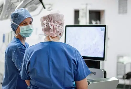
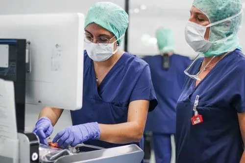
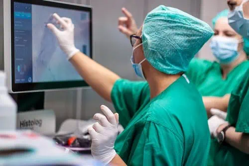
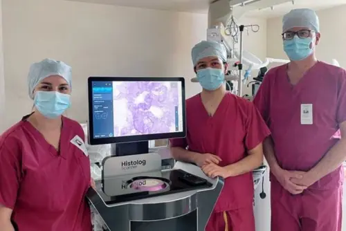

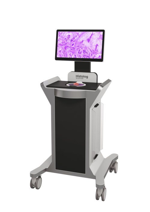
The Histolog® Scanner
Built on ultra-fast confocal microscopy technology
Highly practical for quick assessment at the point-of-care
Bringing live cancer cells to the clinician’s fingertips
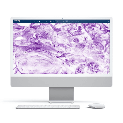
The Histolog® Scanner, developed by SamanTree Medical, represents a significant advancement in medical imaging, particularly in the field of cancer treatment. This state-of-the-art device utilizes ultra-fast confocal microscopy technology to offer high-resolution imaging of the surface of fresh tissue. The technology has been CE marked since 2016, indicating its compliance with health, safety, and environmental protection standards within the European Economic Area.
Europe
SamanTree Medical SA
E40 Business Park
Rue de Bruxelles 174
B-4340 Awans, BELGIUM
+32 4 290 81 63
co*****@*******ee.com
www.samantree.com
United States
SamanTree Medical Inc
50 Milk Street, Floor 16
Boston, MA 02109
866.993.1441
co*****@*******ee.com
www.samantree.com
This document is intended solely for healthcare professionals.
The Histolog Scanner is a CE marked product commercialized in Europe; TGA approved commercialized in Australia and granted 510(k) clearance for commercialization in the US.
Histolog is a registered trademark in the US and internationally for SamanTree Medical SA.
©2025 SamanTree Medical SA. All rights reserved.
DOC number-WW and Version number (E.g. DOC001-WW V.1.0)
CE MARK
Intended use
The Histolog® Scanner is a confocal microscopy device intended for imaging the surface of excised human tissue specimens to visualize morphological microstructures. The Histolog® Scanner is intended to be used in combination with the following accessory devices:
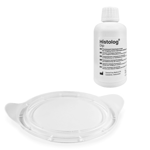
■ The Histolog® Dish, a single-use protection preventing direct contact of the tissue specimen with the Histolog® Scanner during imaging.
■ The Histolog® Dip fluorescent histological stain.
The Histolog® Scanner and its accessory devices are intended to be used in vitro for the examination of specimens. They are in-vitro diagnostic medical devices.
The Histolog® Scanner and its accessory devices are intended to be operated by trained healthcare professionals.
US Important Safety Information

The Histolog® Scanner
Indications for use: The Histolog® Scanner is a confocal laser system intended to allow imaging of the internal microstructure of tissues including, but not limited to, the identification of cells, vessels and their organization or architecture.
Warnings:
The Histolog® Scanner and its accessory devices are intended to be used in vitro for the examination of specimens. They are not intended for use on patients.
■ The Histolog® Scanner and the Histolog® Dish are non sterile devices. For use in an intraoperative setting, the Histolog® Scanner and its accessory products shall be located outside the sterile surgical area.
■ When handling potentially infectious tissue specimens, avoid skin contact by wearing gloves or other protective equipment.
■ This device is not compatible with magnetic resonance (MR) or explosive environments.
■ Handles are not provided for lifting the device, but exclusively have a function for pushing/pulling.
■ The Histolog® Scanner imaging window is fragile. Avoid impact with foreign objects. Do not load with mass above 1 kg. ■ Do not operate the Histolog® Scanner in case of damage to the imaging window.
■ The protective earthing connection of the Histolog® Scanner is performed through the power cord connection. If this cable is damaged, the device must not be connected to the mains power.
Cautions:

■ Only use compatible accessories and parts supplied by SamanTree Medical or by an authorized distributor.
■ Do not re-use a Histolog® Dish. A new Histolog® Dish shall be used for every new surgical procedure.
■ Do not use a Histolog® Dish, if it is damaged or if its optical interface is visibly dirty.
■ If the information about the tissue specimen orientation is critical, the tissue specimen should be marked for orientation before starting the imaging procedure with the Histolog® Scanner.
■ Do not apply tissue marking dyes on the tissue specimen before imaging it with the Histolog® Scanner. Tissue marking dyes prevent proper imaging with the Histolog® Scanner.
■ Do not use disinfecting products that contain glycolic acid. These disinfecting products may damage the Histolog® Scanner.
■ The Histolog® Scanner is intended for use in the electromagnetic environment specified in the Histolog Scanner Instructions for use §16.2 and §16.3.
■ Do not use this device in proximity to sources of strong electromagnetic radiation (e.g. unshielded intentional radio frequency (RF) sources), as these can interfere with proper operation.
■ Do not use this device in proximity of active HF surgical equipment, as these can interfere with proper operation.
Laser safety
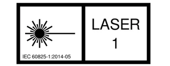
The Histolog® Scanner is a Class 1 laser product, as per IEC 60825-1:2014-05. Laser radiation in the visible wavelength range is emitted from the imaging window [G] during imaging.
The Histolog® Scanner embeds a Class IIIB laser component with the following characteristics:
Wavelength: 488±4 nm
Max output power: 200 mW (CW); Beam divergence: < 0.7 mrad
More Solutions from Saman Tree Medical

Compare Solutions
Get Pathology News Delivered to Your Inbox
Learn More

Saman Tree Medical News

Histolog Scanner Confocal Microscopy: Pathology Image Evaluation of Surgical Margins in Robot Assisted Radical Prostatectomies
Editor2026-05-22T14:39:42+00:00July 9, 2025|

Intraoperative Margin Assessment for Breast Conserving Surgery – the advantages of the Histolog® Scanner
admin@pathologynews.com2026-05-29T15:30:24+00:00May 8, 2025|
Field Service Engineer – Liège, Belgium
admin@pathologynews.com2024-08-12T08:48:40+00:00August 12, 2024|

Supplementary Webinar Q&A
admin@pathologynews.com2024-02-26T11:04:09+00:00February 26, 2024|

Precision in Prostatectomy: The Synergy Between Robot- Assisted Radical Prostatectomy and the Histolog Scanner
admin@pathologynews.com2026-05-22T14:53:16+00:00February 7, 2024|

Reviews
There are no reviews yet.