
TLS Detect
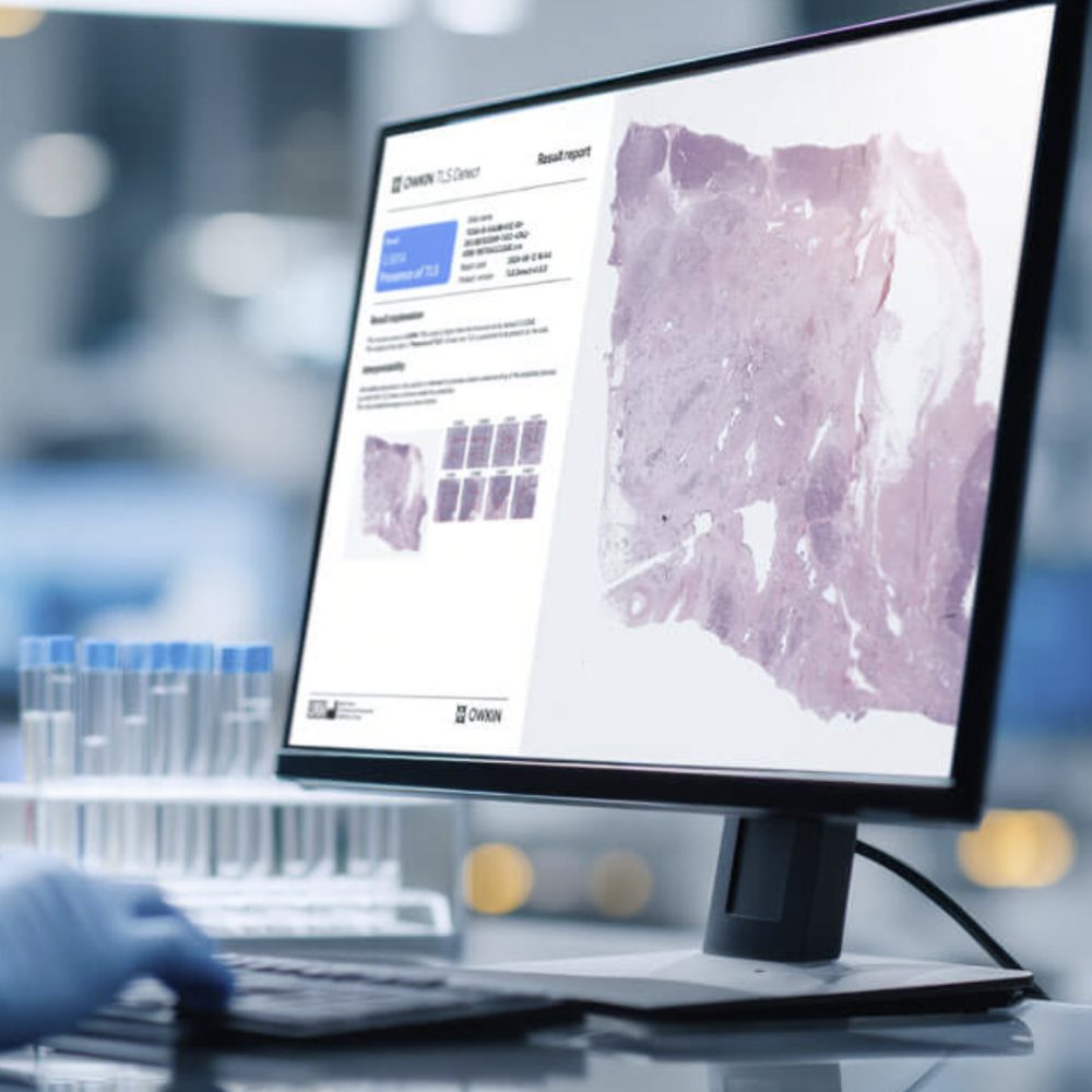

Set the standard for TLS detection
Detect Tertiary Lymphoid Structures across tumor types from digitized H&E slides
Gain insights into a tumor’s interaction with the immune system
TLS provide crucial insights to better understand the response to immunotherapies*
A major player in antitumor immune response, the presence of TLS has been associated with improved survival in several tumor types.1
During drug development and clinical trials TLS can serve as a predictive biomarker for response to certain immunotherapies.
Presence of TLS potentially indicates an enhanced immune response making it a possible target for improved response to immunotherapy.2
TLS could guide the development of therapies that target both the tumor cells and the local immune environment.
TLS can support clinical trial optimization, to help better select trial participants to improve success rates across trial phases.
Gain insights into a tumor’s interaction with the immune system
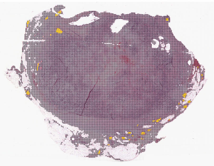
As a novel biomarker, TLS presence helps stratify the overall survival risk of untreated cancer patients and helps identify those who may benefit from efficient immunotherapy treatment.
Waiv’s simple and standardized approach could support the adoption of this biomarker as an independent predictor of response to immune checkpoint inhibitors (ICI).
With a sensitivity of 97%, TLS Detect supports the screening for TLS presence across tumors within a research setting.
An automated solution providing standardized quick results

Suitable for tumor specimens across multiple indications

Works with tumor tissue on digitized H&E slides

Externally validated high sensitivity

Researchers in a non-clinical routine setting

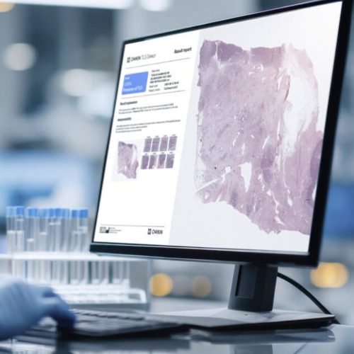
Delivered as a PDF with intuitive design

Notes the presence or absence of TLS with score predictions
More Solutions from Waiv
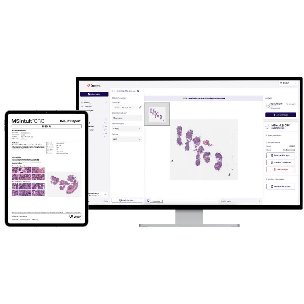
Compare Solutions
Get Pathology News Delivered to Your Inbox
Learn More
Waiv News
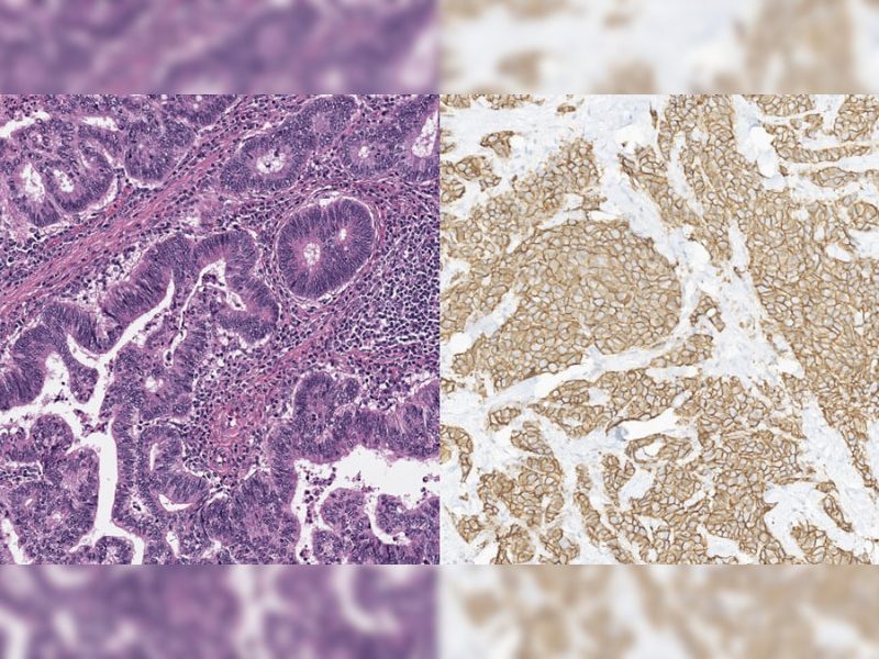
Waiv Enters Collaboration with Daiichi Sankyo to Deliver AI-Derived Biomarkers for ADC Program
Editor2026-05-08T10:36:28+00:00May 7, 2026|
![Figure 1: Fine-tuning improves robustness and performance jointly. Each foundation model is shown before (open circle) and after (filled circle) fine-tuning. The x-axis is the average PathoROB robustness index over three datasets, where higher values indicate greater robustness; the y-axis is the normalized rank sum over the HEST, THUNDER and Patho-Bench benchmarks, rescaled to [0, 1] so that 1 corresponds to the best achievable performance.](https://www.pathologynews.com/wp-content/uploads/2020/07/embedding-figure-800x600-1.png)
Meet Phaet and Mascaret: two pathology foundation models built to generalize across labs
Editor2026-08-10T07:54:33+00:00July 30, 2020|

Reviews
There are no reviews yet.