ARTIFICIAL INTELLIGENCE DECISION-MAKING AND DIAGNOSIS AUTOMATION IN THE PATHOLOGICAL ANATOMY DEPARTMEN
Bicêtre Hospital is a pioneer in the integration of artificial intelligence in pathological anatomy, illustrating the promising future of this technology in the medical field.
With the gradual digitization of pathological anatomy services, the use of artificial intelligence tools for clinical decision support and the automation of certain tasks, under the sovereign control of pathologists, is accelerating.
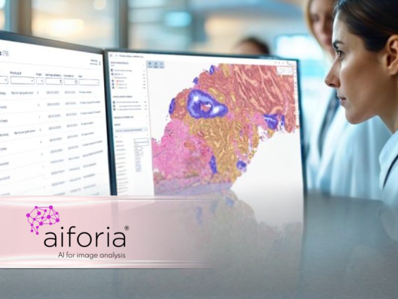
This is precisely what the Finnish company Aiforia Technologies, which specialises in digital image analysis for many types of cancers via Artificial Intelligence and Deep Learning, offers. The Bicêtre hospital has taken the plunge and should bring other AP-HP establishments in its wake.
FACILITATE THE ANALYSIS OF PROSTATE BIOPSIES:
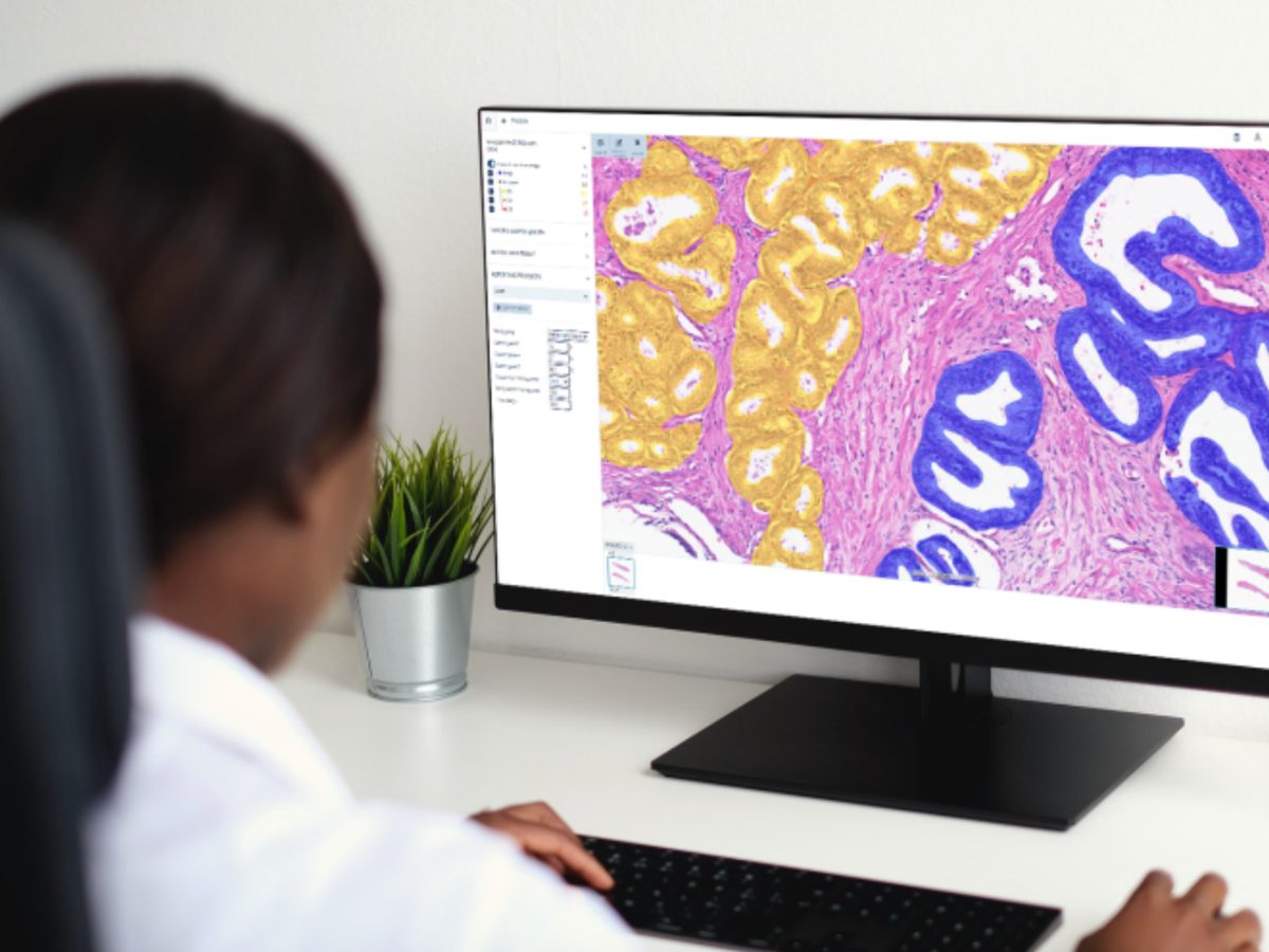
At the Bicêtre hospital, last year, nearly 7,000 prostate biopsy slides were treated. The analysis of these slides, complex and time-consuming, was quickly identified as an appropriate area for the deployment of a decision support tool. “We are facing a real shortage of pathologists,” says Prof. Sophie FERLICOT, head of the pathological anatomy and cytology department. At the same time, given the number of biopsy slides to be analyzed for a patient (minimum of 24 slides) and the size of the foci to be studied, AI quickly became a logical next step in the digitization of our department. Especially since many of the steps in this analysis are long, tedious and repetitive: measurement of prostate tissue, measurement of the cancerous focus, evaluation according to the ISUP/Gleason score, etc.
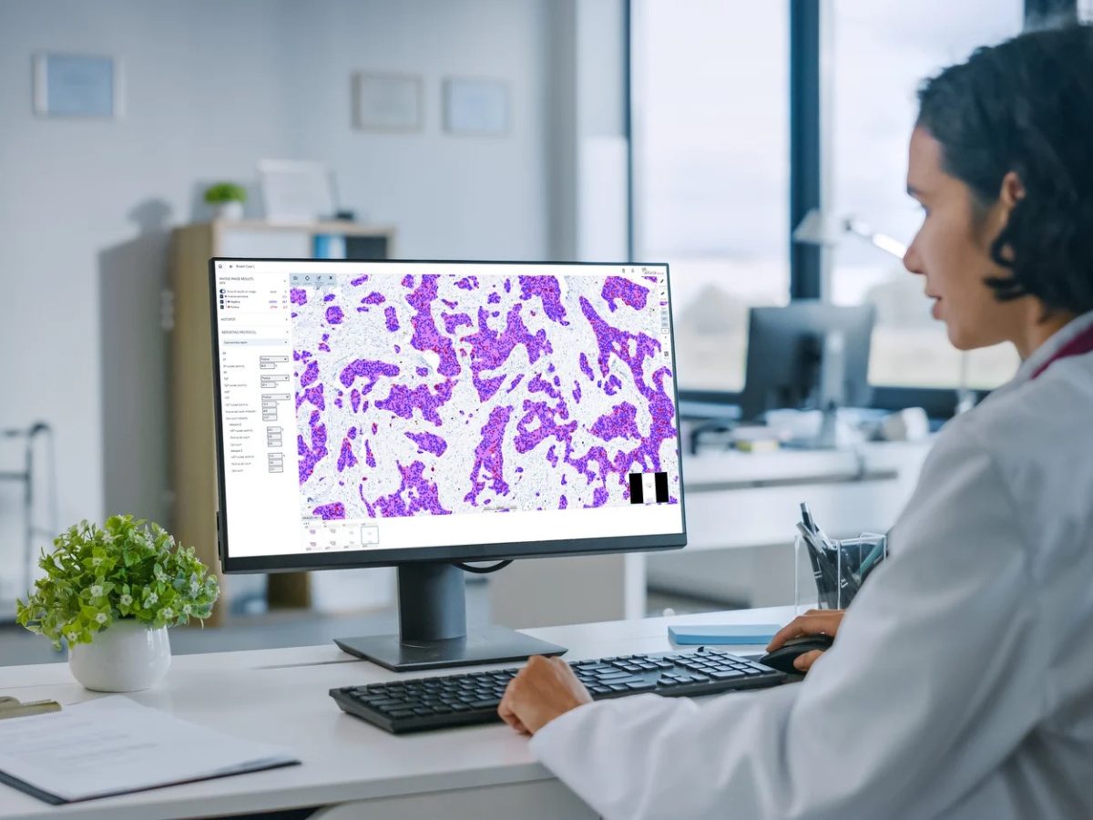
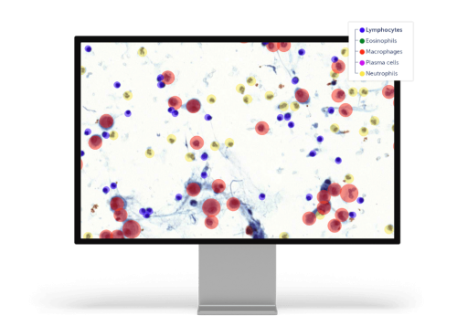
After a thorough evaluation, the company Aiforia Technologies was chosen. The Aiforia® Clinical Platform is an innovative cloud-based solution offering AI-assisted image analysis suites, suitable for the diagnosis of multiple cancers (prostate, breast, lung, stomach, etc.). It also offers advanced visualization, assisted screening, and reporting tools, facilitating medical care.
The results were quickly medically validated.
“We benefit from better cancer detection. The average rate of failure to detect cancer foci has been estimated at 7% (Lancet Oncology). It goes to 3% with AI. Another advantage is that with more precise detection, we have less use of immunohistochemical analyses,”
says Dr. Clovis Adam, a hospital practitioner in Pathological Anatomy in Bicêtre.
INTEGRATION WITH THE WORKFLOW:
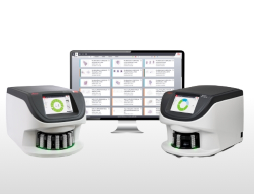
The solution has been integrated into the entire laboratory ecosystem: slide scanners, SGI (Tribune Health Image Management System).
“We have worked closely with third-party vendors to maximize the effectiveness of our tools. With this synergy, we have been able to automate many key processes, which not only speeds up repetitive tasks but also improves the accuracy and consistency of analyses. This approach meets the needs of laboratories in terms of efficiency and productivity, while ensuring a high level of diagnostic quality,”
explains Julien Courtial, sales manager at Aiforia Technologies.
Thus, some measurements can be made by the tool.
“However, the user interface allows us to make corrections very easily if necessary. The algorithm instantly updates the analysis by incorporating these adjustments, ensuring optimized results without wasting time. The solution offers great flexibility to adjust and challenge the model, while allowing, if necessary, to request additional examinations to refine the diagnosis,”
insists Prof. Sophie Ferlicot.
“It is essential that the pathologist remains in control of the interpretation,”
emphasizes Professor Catherine Guettier.
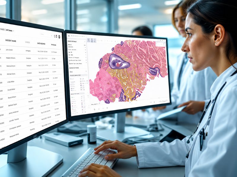
Thus integrated into the laboratory ecosystem, the tool also organizes the workflow, making it possible to prioritize the most serious cases. The benefits for patients are obvious. Less time per slide means more doctor’s time, with a more detailed and faster analysis. The deployment of Aiforia at AP-HP should therefore continue to extend to the 6 departments performing analyses of digitized prostate slides.
Share this article




Follow us
Get Pathology News Delivered to Your Inbox