Flash Scanner FS4-R

Flash pathology logo

Instant pathology – No tissue preparation, no labelling, no slicing

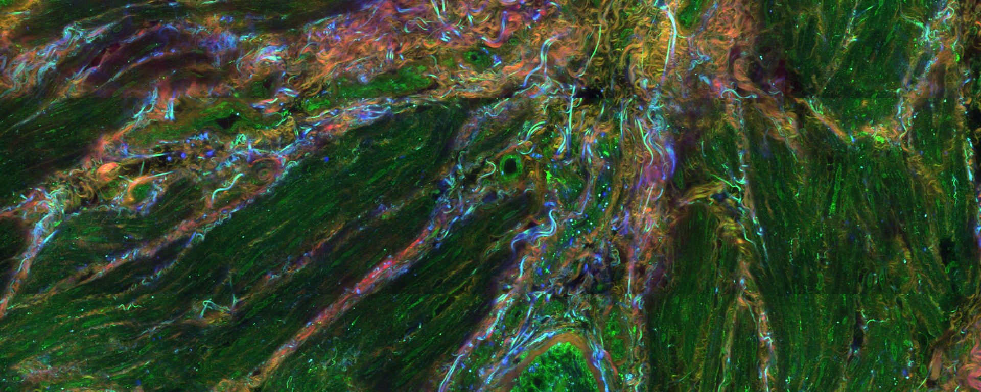
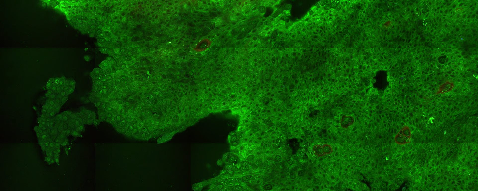
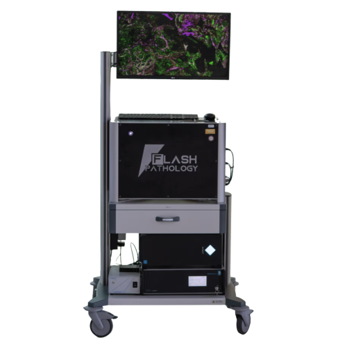
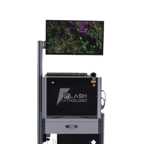
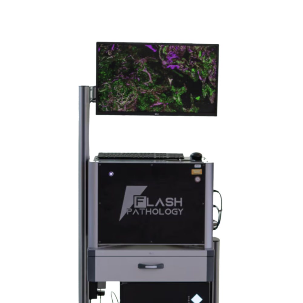
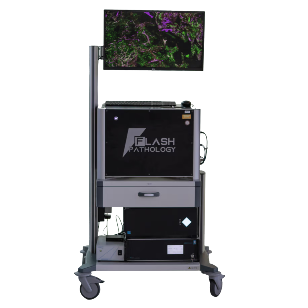
The Flash Scanner FS4-R is a state-of-the-art microscope that delivers rapid, label-free, subcellular 2D and 3D imaging of fresh unprocessed tissue. By integrating higher harmonic generation (HHG) with multiphoton fluorescence techniques, it provides real-time insight into tissue architecture and cellular morphology, without preparation or post-processing, leaving samples intact for advanced downstream analysis. Designed as a transportable system, it can be easily moved between laboratories or clinical settings. With its unique combination of speed, rich histological insights, non-destructive imaging, and zero sample preparation, the FS4-R paves the way for intra-operative biopsy adequacy assessment, tissue pre-selection for advanced analysis, and cell dynamics studies of drug responses.
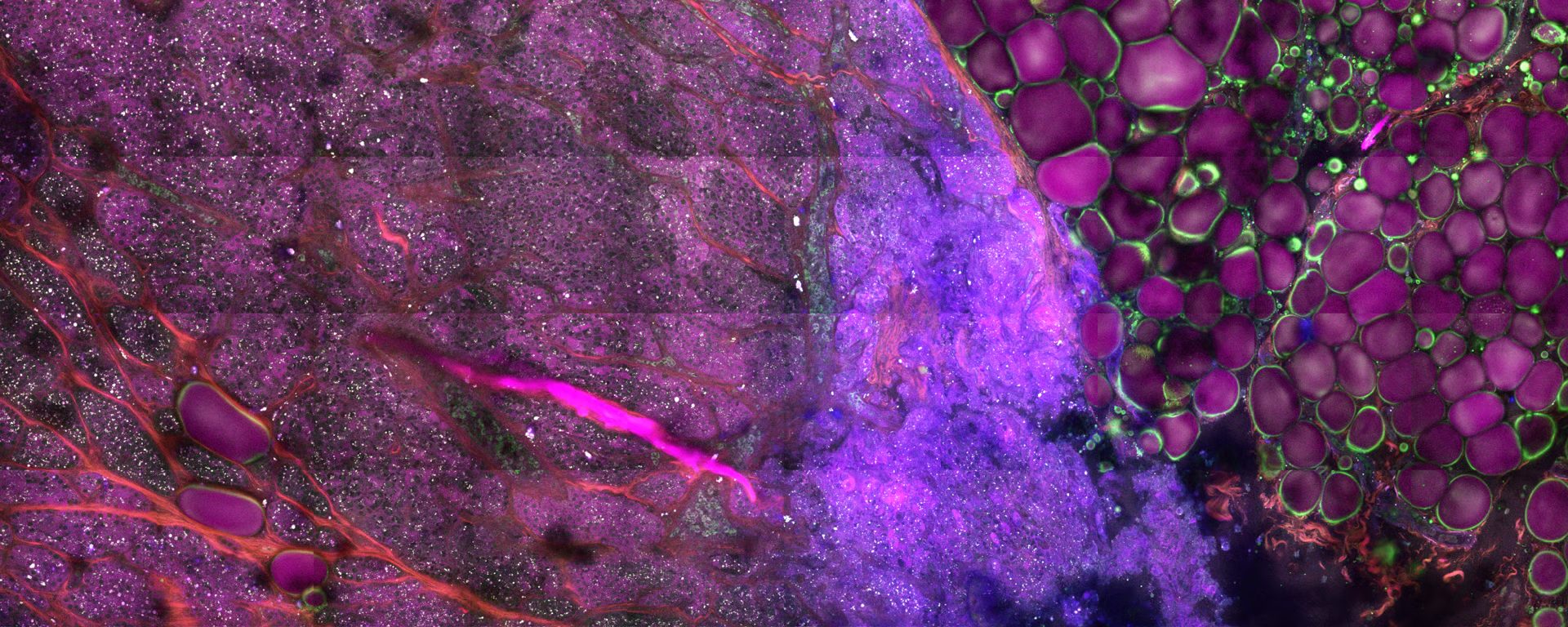


>> Subcellular resolution

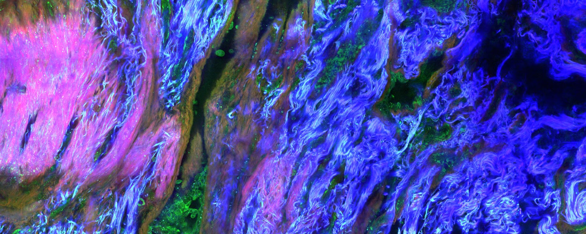
Reveal key cellular and histological hallmarks using four distinct imaging modalities.

>> Label-free imaging

Visualize tissues without the use of labels or dyes, preserving samples for further analysis.

>> 3D tissue visualization

Generate 3D images using automatic optical sectioning – no sample slicing required.

>> Quick turnaround

Minimize specimen preparation time and evaluate your samples in just minutes.

>> Publication-ready images

Capture vibrant, high-quality images that effectively convey the story behind your data.

How does the Flash Scanner FS4-R work?

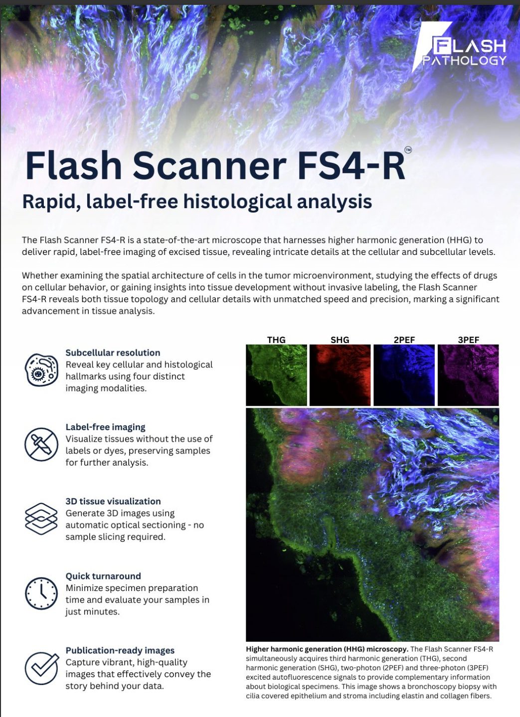
The Flash Scanner FS4-R is an imaging platform designed for rapid, label-free histological assessment of unprocessed tissue samples. The functionality of the Flash Scanner is based on the detection of nonlinear multiphoton signals, including higher harmonic generation (HHG) signals, such as second and third harmonic generation (SHG and THG), as well as two-photon and three-photon excited autofluorescence (2PEF and 3PEF). These imaging modalities provide complementary information on both tissue architecture and cell morphology, allowing to successfully discriminate between different types of tissues.

What tissue structures can be visualized with the Flash Scanner?

The Flash Scanner FS4-R offers four unique imaging modalities:

  • SHG: Captures non-centrosymmetric molecules and structures, like collagen fibers, microtubules, and myofilaments.

  • THG: Visualizes cellular structures with distinct refractive indices, such as cell membranes, lipid droplets, and myelin sheaths.

  • 2PEF and 3PEF: Detect the presence of intracellular molecules, such as flavins, NAD(P)H, retinol, and tryptophan, as well as extracellular structures, including elastin and collagen fibers.

Can the tissue samples imaged with the Flash Scanner FS4-R be used for further analysis?

Yes, the tissue samples imaged with the Flash Scanner FS4-R can be used for further analyses, such as histology, molecular studies, or other imaging techniques. The Flash Scanner utilizes non-invasive, label-free imaging modalities, such as higher harmonic generation and multiphoton excited autofluorescence, therefore the samples remain intact and undamaged.

Can the Flash Scanner FS4-R image tissue samples without any preparation or processing?

Yes, the Flash Scanner FS4-R can image fresh tissue samples without the need for staining, labeling, or other forms of tissue processing. Higher harmonic generation directly utilizes the optical properties of the tissue, allowing for high-resolution imaging while preserving the sample’s natural structure and composition.

Screenshot 2025 09 12 142908
Optical Resolution

0.4 µm (lateral), < 3 µm (axial)

Field of view (single image)

400 µm × 400 µm

Max. Mapped Field

Ø = 20 mm (limited by the size of the sample dish, larger scan range on request)

Scanning Speed

2 Mpixels/sec

Imaging Modes

Inspection mode (1 pixel/µm) and high-quality mode (5 pixels/µm)

Imaging Wavelength

1070 nm

Operating Temperature and Electrical Requirements

18˚C to 30˚C; 110 – 230 VAC, 50 – 60 Hz

Deployment Environment

Research Only

Regulatory Certification

CE

More Solutions from Flash Pathology

  •  flash pathology product

Get Pathology News Delivered to Your Inbox

Learn More

flash pathology square logo

Flash Pathology News

Flash Pathology Solutions