Consisting of a pathology viewer and a server and storage application, Philips Image Management System (IMS) is designed to enhance the efficiency and effectiveness of your pathology lab. The open, scalable design and new user interface integrate into your workflow and IT infrastructure environment. The IMS manages image repositories – including whole slide images (WSI) and gross images – and comprehensive LIS integration, networking, security, audit trail and archiving capabilities.
The intelligent backbone of your digital pathology workflow
The Philips Image Management System (IMS) is the central hub of Philips IntelliSite Pathology Solution (PIPS), uniting whole‑slide images, clinical data, and advanced applications in one workspace.
Built on a modern, web‑based architecture, IMS integrates with your IT environment and scales from single‑site to multi‑site networks. By bringing images, case information, and collaboration tools together, it enhances the speed, consistency, and confidence of diagnostic review.
Fast, scalable Image Management
At the core of IMS is a powerful image repository that securely stores, organizes, and manages whole-slide images and associated data. Its open and modular design supports efficient and reliable access to images, even at high volume, enabling laboratories to scale confidently as workloads grow. Pathology image management can be tailored to meet diverse needs, whether through a customizable on-premise storage configuration or in combination with secure cloud-based archiving. PIPS offers robust repository management with LIS interoperability, network flexibility, and integration with existing systems. Cloud Data Services facilitates scalable, cost-effective storage with instant access, and a foundation for AI-powered integrated diagnostics.1
A cloud‑enabled deployment reduces reliance on on‑premise infrastructure while providing the flexibility, performance, and scalability required for enterprise collaboration and AI integration. This represents an important next step in advancing our digital pathology strategy.
– Dr. Cordon-Cardo, Chair of the Department of Pathology, Molecular and Cell-Based Medicine at Mount Sinai Health System
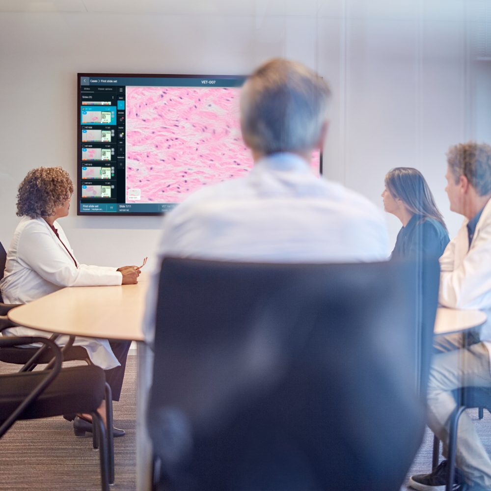
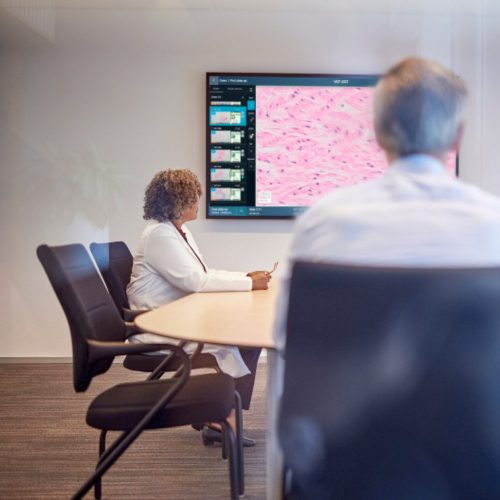
The IMS viewer is designed to help pathologists work faster and more intuitively, offering fluid navigation, smart caseload management, and real-time collaboration tools. By enabling teams to share cases, whether for tumor boards, second opinions or routine consults, helps to minimize workflow interruptions and supports a more predictable and efficient diagnostic process.
Independent evaluations reinforce these benefits. In the KLAS Digital Pathology report2, users highlighted faster turnaround times, improved workflow reliability, and reduced case handling steps thanks to streamlined image access and integrated case information. Likewise, the Granada study demonstrated significant efficiency gains of up to 25%3 when using digital workflows supported by IMS, driven by smoother case navigation, reduced slide handling, and improved collaboration across sites.
LIS and Enterprise interoperability for connected workflows
Because pathology workflows rely on fully connected systems, IMS integrates deeply with your laboratory information system (LIS) and broader enterprise infrastructure. Features such as contextual IMS launch from LIS worklists and synchronized case metadata, reduce manual data entry and reduce turnaround delays.
AI interoperability to enhance accuracy and efficiency
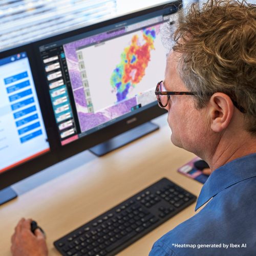
IMS is engineered as an open platform for AI4, making it possible to incorporate advanced algorithms directly into the viewer experience. By connecting to third-party AI applications including contextual launch of Ibex AI5 directly from the IMS, Philips digital pathology solution helps pathologists gain AI-driven insights without exporting data or disrupting workflow.
This open AI strategy empowers sites to choose the algorithms that best fit their needs today, while maintaining flexibility to integrate future innovations. In real‑world clinical use, this approach has translated into substantial performance gains, including a 43‑hour improvement in turnaround time when using Ibex AI with PIPS (reducing TAT from 1.8 days to just 9.4 minutes) and productivity improvements of up to 37%,6 demonstrating how tightly integrated AI can meaningfully accelerate diagnostic workflows.
Standards based open ecosystem
Philips actively supports interoperability with viewers, scanners, PACS, VNAs and AI tools from multiple vendors.4 This standards-based approach protects your investment and enables cross-modality collaboration across the broader health system. Combined with a scalable architecture and integration into enterprise IT, IMS provides a foundation for digital pathology that grows with your clinical, operational and technological needs.
Pathology Scanners
Image Management System
End-to-end Services
1 Third party claims from AWS, which may not apply in all markets and may be updated from time to time. The functionalities and benefits of the solution depend on customer-specific configuration and use. Please contact your local Philips representative for (market) availability.







Reviews
There are no reviews yet.