Prostate cancer is among the most diagnosed cancers in men, affecting approximately one in eight men during their lifetime.1 At the same time, prostate cancer is not a single disease but a broad spectrum of conditions with widely varying clinical behavior.
Overstaging and understaging
Diagnosing prostate cancer is not always straightforward. The key challenge lies in accurately distinguishing between slow-growing cancers that may never cause harm and more aggressive forms of the disease that require timely treatment, ensuring that patients receive the right treatment at the right time. Despite advances in radiology and pathology, diagnostic variability remains a significant challenge in prostate cancer care. Understaging can delay or limit treatment of aggressive disease, leading to progression of the disease, higher downstream costs, and poorer patient outcomes.2 Overstaging can drive unnecessary radical treatments, exposing patients to avoidable side effects and increasing long-term cost of care.2
The hidden cost of negative biopsies
Standard systematic prostate biopsies often yield negative results, confirming no cancer is present. These procedures place a significant burden on urology services and pathology departments, consuming procedure room capacity, clinician and staff time, diagnostic resources and follow-up consultations without delivering clinical benefit.3 At scale, unnecessary biopsies represent a substantial and often underrecognized financial and operational burden, particularly for pathology departments and laboratories that are already overburdened and facing workforce shortages.3
Improving diagnostic efficiency and confidence across radiology, urology and pathology
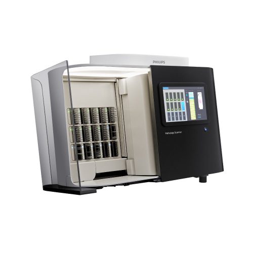
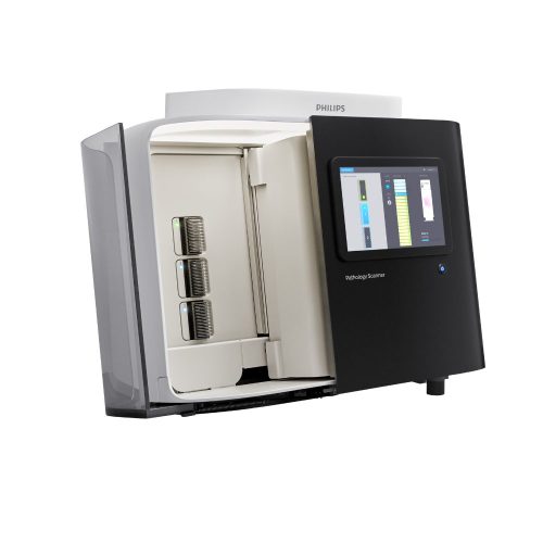
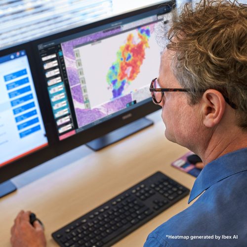
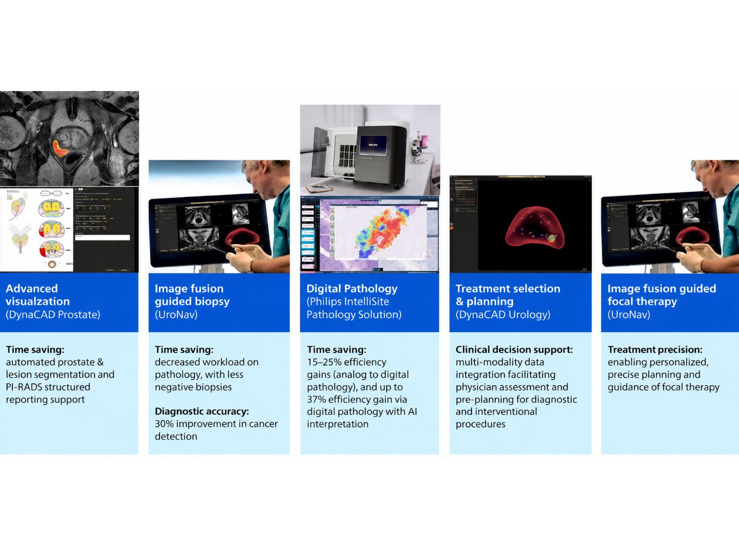
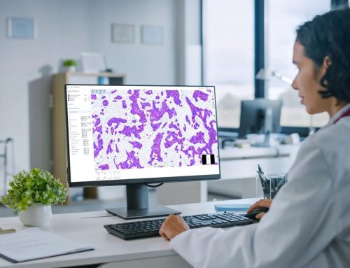
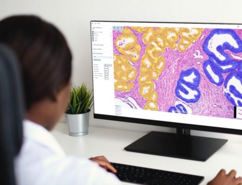
Philips DynaCAD Prostate with Quibim AI-powered*,4,10 automation improves radiology workflow efficiencies, while UroNav enables MRI–ultrasound fusion-guided biopsy. As a result, hospitals achieve 30% higher detection rates of clinically significant cancer, reduced detection of insignificant disease, fewer biopsy cores, and lower rates of repeat and negative biopsies.5 The tissue samples are subsequently processed and analyzed by the pathologist to establish a definitive diagnosis. Pathology remains the definitive step in prostate cancer diagnosis, yet pathology departments face increasing pressure from rising case volumes and workforce constraints. Philips IntelliSite Pathology Solution addresses these challenges by enabling fully digital workflows that support remote reading, improved collaboration, and more efficient workload distribution and pathology reading, helping departments maintain or even shorten turnaround times while scaling services. The transition from analog to digital pathology alone has been shown to deliver efficiency gains of approximately 15–25%,6 while the addition of advanced AI tools can further enhance productivity. Ibex AI9 contextual launch further strengthens diagnostic performance by augmenting the pathologist’s expertise.7 By identifying areas suspicious for cancer, supporting Gleason grading, and providing quantitative insights, Ibex AI improves consistency and reduces inter-observer variability,7 with overall efficiency gains reported of up to 37%.8 Together, Philips digital pathology and Ibex AI9 allow hospitals to deliver confident, timely diagnoses7,8 while making more efficient use of specialized pathology resources.
From integrated data to targeted therapy: enabling treatment selection, planning, and guidance
By integrating radiology, urology, and pathology data within DynaCAD Urology, clinicians gain a unified view of each patient’s case, supporting personalized treatment decisions. For appropriately selected patients with localized, clinically significant prostate cancer, focal therapy represents an important evolution in treatment strategy. Rather than treating the entire prostate gland, focal therapy targets only cancerous tissue, preserving healthy structures whenever possible. DynaCAD Urology and UroNav enable personalized and precise planning and guidance of focal therapy using the same image guided targeting principles applied in biopsy procedures. This precision allows clinicians to effectively treat cancer while minimizing damage to surrounding tissue.
Advancing value-based prostate cancer care
As healthcare systems continue their shift toward value-based care, prostate cancer represents a critical opportunity to improve outcomes while controlling costs. Precision diagnostics, AI-enabled workflow automation, and targeted therapies allow hospitals to move beyond one-size-fits-all approaches toward more efficient and personalized care models.
References
- https://www.cancer.org/research/acs-research-news/prostate-cancer-is number-1-for-118-countries-worldwide.html
- https://doi.org/10.3390/diseases13060167
- https://doi.org/10.1016/j.jval.2021.07.002
- https://quibim.com/qp-prostate/
- Siddiqui MM, et al. Comparison of MR/ultrasound fusion–guided biopsy with ultrasound-guided biopsy for the diagnosis of prostate cancer. JAMA. 2015;313(4):390-397
- KLAS Research ‘US Digital Pathology 2023’ Performance Insights
- Information of Ibex AI. https://ibex-ai.com/resource/prostate-cancer-ai pathologist-used-to-help-diagnosis/
- Information provided by Ibex AI: Raoux, et al. Modern Pathology (2021) 34 (suppl 2): 598-599. https://www.youtube.com/watch?v=9XuWK8PmUlw
- Ibex AI is Research Use Only in the United States
- Quibim QP-Prostate is only available in USA
Share this article



Follow us
Get Pathology News Delivered to Your Inbox