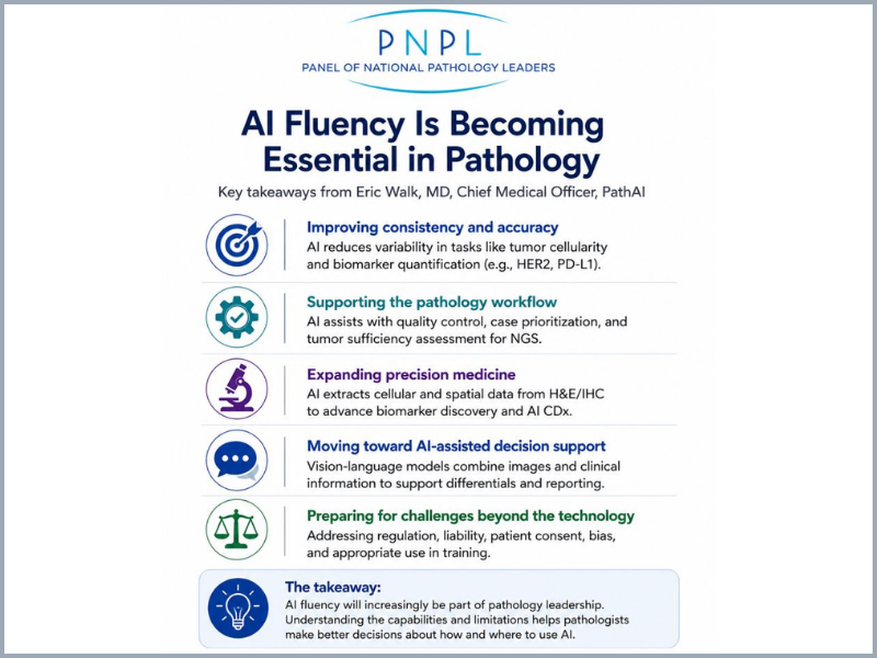
Artificial intelligence is moving quickly into pathology, but Dr. Eric Walk, Chief Medical Officer of PathAI, believes adoption is getting ahead of understanding. During PNPL’s Q3 Digital Pathology & AI Focus Group meeting on July 24, 2026, he urged pathology leaders to make AI fluency a priority – not so pathologists can build algorithms themselves, but so they understand enough about the technology to determine which tools are appropriate for their practices.
Dr. Walk noted that pathologists routinely understand the basic science behind technologies they use every day, including immunohistochemistry (IHC), PCR, and next-generation sequencing. Yet many could not explain, even at a high level, how a neural network works. As AI becomes part of diagnosis, biomarker assessment, workflow, and precision medicine, he argued that this gap matters. Without a basic understanding of how an AI model was developed and trained, pathologists may have difficulty evaluating whether it is suitable for a particular clinical use.
His presentation covered four areas: (1) basic AI and machine-learning concepts; (2) how different methods apply to clinical pathology and precision medicine; (3) emerging uses of generative AI and vision language models; and (4) the technical, regulatory, ethical, and medical-legal issues that remain unresolved.
What Pathologists Need to Know About AI
Dr. Walk began by encouraging pathologists to be more precise when talking about AI. “Artificial intelligence” is a broad umbrella that includes machine learning, deep learning, and generative AI, each with different capabilities. Most current pathology applications use deep learning, in which neural networks learn patterns from data and make predictions, such as distinguishing tumor from non-tumor tissue or quantifying a biomarker. Generative AI, which includes large language models, is beginning to enter pathology but currently represents a smaller portion of clinical applications.
He used a simple image-classification example to explain how neural networks are trained. An image is divided into smaller patches, which are processed through layers of a network. During training, the model repeatedly adjusts internal parameters as it learns from examples; once trained, it can apply what it has learned to new images, a process known as inference. The same basic principle can be applied to a whole-slide pathology image, where the output might identify tumor, necrosis, normal tissue, or another feature.
Another increasingly important concept is the foundation model. Rather than being a single model that can perform every pathology task, a foundation model is a large model trained on broad datasets that serves as a backbone for more specific applications. It can then be adapted for tasks such as tumor detection, H&E diagnosis, PD-L1 scoring, or tissue segmentation. As illustrated in Dr. Walk’s slides, the advantage is that a foundation model has already learned a broad representation of histopathology, reducing the amount of additional data needed to develop individual applications.
The practical lesson for pathology leaders is that the method should fit the application. A model designed to count PD-L1-positive cells, for example, should not necessarily be built the same way as a model looking for previously unknown morphologic patterns associated with a genetic mutation. Understanding these distinctions helps pathologists ask better questions when evaluating a product rather than relying solely on claims that it “uses AI.” The slide deck summarizes the differences among supervised, weakly supervised, moderately supervised, and generative approaches, along with their advantages and limitations.
Digital Pathology, Workflow, and Clinical Applications
Dr. Walk next placed AI within the broader digital pathology environment. A digital system still depends on the laboratory information system, image management system, scanners, storage, and interfaces that connect them. AI can function separately, but he strongly favored close integration between AI and the image management system because the value of an algorithm can be lost if using it requires cumbersome extra steps.
This point became especially important during the discussion. One PNPL participant described evaluating prostate cancer AI tools that performed well technically but were too difficult to incorporate into daily sign-out. Dr. Walk recommended evaluating not only the quality of the algorithm but also the user interface, number of steps required to use it, and the broader AI ecosystem supported by the image management system. Choosing a platform that works well today but limits access to useful third-party AI applications later could restrict a laboratory’s options.
AI also has potential upstream of the pathologist. Dr. Walk described possible applications in slide quality control, case prioritization, workload distribution, tumor identification, cell counting, and biomarker quantification. Instead of assigning cases based only on number, for example, AI could help account for complexity so workloads are distributed more evenly.
These applications are increasingly relevant as case volumes and complexity rise. Even where the absolute number of pathologists is relatively stable, Dr. Walk noted that many pathologists report increasing workloads and more time required per case, particularly as biomarker assessment becomes more complex. In that environment, the case for AI is not simply that a computer can perform a task; it is that technology may help laboratories use limited professional time more effectively.
Precision Medicine and More Reproducible Biomarkers
A major portion of the presentation focused on precision medicine, where Dr. Walk sees significant potential for computational pathology.
Many biomarkers used to determine eligibility for cancer therapies depend on visual assessment by pathologists. HER2 and PD-L1 are familiar examples, but scoring can vary among observers, particularly around clinically important cutoffs. PD-L1 scoring can theoretically require cell counting, although in practice pathologists often estimate the proportion of positive cells.
AI can approach these tasks differently by identifying tumor and counting individual cells, producing a quantitative result rather than an estimate. Dr. Walk reviewed studies in which AI-assisted assessment improved reproducibility and described research comparing AI results with expert pathologists and clinical outcomes.
He also discussed a broader change underway in drug development: using AI to extract information from routine H&E and IHC slides that humans cannot reliably measure at scale. Machine learning can quantify cell numbers, staining intensity, tissue areas, and spatial relationships between different cell populations, turning morphology into structured data that can be searched for potential biomarkers.
This may be especially useful in developing new cancer therapies. Rather than beginning with a predefined biomarker and asking whether it predicts response, researchers can use machine learning to search for previously unrecognized morphologic patterns associated with outcomes. Dr. Walk described this as a shift from purely hypothesis-driven biomarker development toward hypothesis-seeking discovery.
The approach brings an important limitation, however. During the discussion, Dr. David Klimstra, Co-Chair of PNPL’s DP & AI Focus Group and former Chief Medical Officer at Paige AI, noted that the strongest AI predictor may eventually be something a human pathologist cannot readily recognize or explain. Dr. Walk agreed that explainability remains a challenge. A model might discover a highly predictive pattern that is difficult to translate into a familiar histologic feature, requiring the field to balance predictive performance with the need to understand how a result was reached.
Decision Support and the Next Generation of AI
Dr. Walk sees generative AI and vision language models (VLMs) as another important area of development. Unlike traditional language models, VLMs can work with both images and text, making them particularly relevant to pathology.
He demonstrated how a pathology-focused chatbot could review an image, describe its histologic appearance, incorporate clinical history, develop a differential diagnosis, suggest appropriate IHC stains, and then reconsider the diagnosis after receiving the staining results. Such tools could be especially valuable for general pathologists who occasionally encounter difficult subspecialty cases outside their usual areas of practice.
The larger opportunity, however, may be pathology decision support. Pathologists must keep track of changing guidelines, new biomarkers, treatment-related testing requirements, prior results, and increasingly complex clinical information. An AI-enabled system could bring relevant information into the workflow when it is needed – for example, alerting a pathologist that a newly approved biomarker applies to the case currently being reviewed.
Dr. Walk also raised the possibility of using generative AI to make pathology reports more understandable to patients. Patients increasingly have direct access to their reports, but the terminology is often difficult to interpret. AI could potentially generate a patient-friendly explanation while preserving the formal diagnostic report for the clinical record.
Important Questions Remain
Despite his enthusiasm about the technology, Dr. Walk emphasized that AI pathology still faces substantial technical, regulatory, ethical, and medical-legal challenges. Even strong models can produce false-positive and false-negative results, particularly when they encounter unusual cases that were not adequately represented in their training data.
Regulation is also evolving. Digital pathology systems have traditionally been cleared around specific combinations of scanners, image management systems, and displays, which can make seemingly routine technology changes complicated. New regulatory approaches may provide developers with more flexibility, but laboratories still need to understand how changes to their digital systems affect validation and compliance.
There are also unresolved questions about patients’ rights and physicians’ responsibilities. Should patients be told when AI contributes to diagnosis or treatment selection? Should they be able to decline its use? What happens when a physician and an AI system reach different conclusions? Who is responsible when an AI-assisted decision causes harm? Dr. Walk presented these as questions the field must address rather than issues with simple answers.
The discussion ended with another concern: training the next generation of pathologists. If residents have unrestricted access to powerful diagnostic AI, there is a risk that they could become dependent on it before mastering basic morphology. Dr. Walk said training programs will need to protect foundational learning, including limiting AI access when appropriate. The goal should be to produce pathologists who can use AI effectively because they understand pathology—not pathologists who require AI to practice it.
Key Takeaways
- AI fluency is becoming a basic leadership skill. Pathologists do not need to develop algorithms, but they should understand enough about how they work to evaluate their suitability for clinical use.
- Be specific about the technology. Machine learning, deep learning, generative AI, foundation models, and vision language models have different purposes and limitations.
- Match the method to the problem. The appropriate AI approach depends on whether the goal is cell counting, diagnosis, biomarker discovery, spatial analysis, or decision support.
- Workflow matters as much as algorithm performance. AI must fit smoothly into the image management and laboratory environment to provide value in routine practice.
- Precision medicine is a major opportunity. AI can improve biomarker quantification and may uncover clinically important information in H&E and IHC slides that cannot be consistently measured by humans.
- Generative AI may expand AI beyond image analysis. Vision language models and other tools could assist with differential diagnosis, reporting, guideline awareness, and patient communication.
- Human oversight remains essential. False results, bias, explainability, regulatory requirements, liability, and patient consent remain important concerns.
- Training programs need to balance AI access with foundational education. Residents must first develop strong diagnostic skills so that AI remains a tool that supports—not substitutes for—their expertise.
Dr. Walk’s central message was straightforward: pathologists do not need to become AI experts, but they do need to become AI-fluent. As laboratories move from digital pathology into increasingly AI-enabled practice, understanding the technology will help pathology leaders choose tools more carefully, integrate them more effectively, and determine where they can provide meaningful clinical value.
Panel of National Pathology Leaders’ Member Practices have access to a wealth of information and tools to help navigate management challenges, including exclusive access to reports, data, and surveys. For information on how to join PNPL or purchase our new 2026 Hospital Negotiations Factbook, see our website or email ak****@*********rs.org.