by Christos Evangelou, MSc, PhD – Medical Writer and Editor
A new study combining multiplex imaging with artificial intelligence (AI) has identified spatial patterns within melanoma tumors that could help clinicians predict which patients are likely to respond to immunotherapy. According to lead investigator Paolo Antonio Ascierto, the study demonstrates that the physical location and interactions between immune cells, and not just their presence, may help better predict treatment outcomes.
The study was presented as a poster at the 2025 ASCO Annual Meeting.
Need for Accurate Response Prediction Tools
Despite advances in melanoma treatment using immune checkpoint inhibitors, some patients do not respond to immunotherapy. Current biomarkers, such as PD-L1 expression and tumor mutational burden, provide a limited view of tumors, which are inherently heterogeneous. Additionally, traditional prediction methods that focus on individual biomarkers overlook the complex spatial relationships between different cell types within the tumor microenvironment.
More accurate prediction tools are needed so that patients who are unlikely to respond to immunotherapy can be spared unnecessary toxicity and offered more effective treatments, while those likely to benefit can receive immunotherapy with greater confidence.
Approach: Imaging Technology Meets AI Analysis
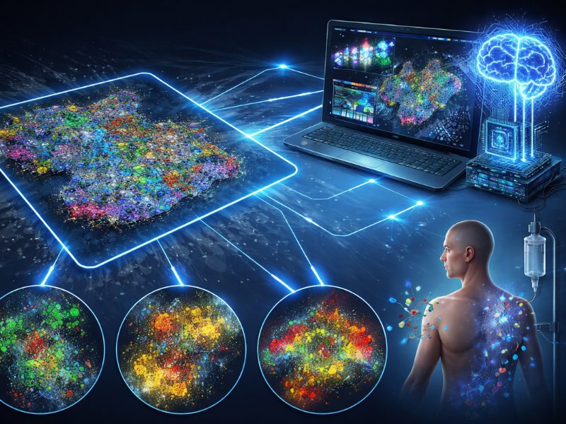
The research team developed a 28-plex panel using sequential immunofluorescence on the COMET platform, which targeted biomarkers associated with tumor microenvironment, immune cell infiltration, and immune checkpoint pathways. According to Dr. Ascierto, this approach represents a significant technological shift from traditional immunohistochemistry.
“Moving from traditional immunohistochemistry to a high-plex platform like COMET was a big step,” Dr. Ascierto stated. “The main challenge was technical: handling 28 markers without compromising tissue quality or signal clarity.”
The researchers analyzed pretreatment biopsies from 12 patients with known long-term response or rapid progression to immunotherapy combination treatment from the SECOMBIT trial. Using Nucleai’s deep-learning-based analysis pipeline, researchers identified 15 cell types, including 10 immune cell populations, as well as 10 cell state markers.
The AI component proved crucial for handling the complexity of the spatial data, according to Dr. Ascierto.
“Another big part was making sure that the image data could be reliably interpreted; this is where Nucleai’s AI pipeline really helped, by automating cell identification and spatial mapping,” he explained.
Spatial Patterns Reveal Treatment Predictors
The pipeline achieved high accuracy, with accuracy and F1 scores exceeding 0.8 for most cell types and markers. The method enabled the quantification of known biomarkers, including T cell activation states, infiltration patterns, and the maturation of tertiary lymphoid structures.
The team compared spatial features between long-term responders and those with rapid disease progression. Interactions between tumor cells, cytotoxic CD8+ T cells, and antigen-presenting cells (APCs) within the tumor were associated with better outcomes. This suggests that successful immunotherapy requires coordinated communication between immune cells directly within the tumor tissue.
Conversely, a high percentage of proliferating regulatory T cells within the tumor invasive margin correlated with worse outcomes. These immunosuppressive cells may create barriers to effective immune responses when concentrated at the tumor boundary, Dr. Ascierto explained.
The tumor microenvironment contained additional predictive patterns. Interactions between endothelial cells and T cells, as well as macrophage proliferation, were associated with immunotherapy resistance. In contrast, interactions between HLA-DR-expressing macrophages and APC cells were associated with treatment response.
Clinical Implications and Future Applications
Dr. Ascierto emphasized that these spatial biomarkers could inform clinical decision-making by providing objective, quantitative measures of immune activity within specific tumor regions. Rather than relying on overall biomarker expression, clinicians could assess the functional organization of immune responses.
"Our hope is to use these spatial signatures to guide treatment decisions, for example, choosing immune checkpoint inhibition as a first-line therapy if we see strong immune activation in the tumor invasive margin," Dr. Ascierto noted. "We're planning prospective studies to validate these signals and refine their role in treatment algorithms."
He added that their approach could also help researchers better understand resistance mechanisms and develop strategies to address resistance by identifying spatial patterns associated with treatment failure.
Potential Implementation Challenges
Dr. Ascierto acknowledged that several challenges need to be addressed before clinical implementation of their method.
“Reproducibility and standardization are the big hurdles,” he said. “Different laboratories use different protocols, and even slight changes in sample handling can affect spatial data.”
Poor sample quality is another issue.
“Pretreatment biopsies can be tricky. Some samples have limited tumor content or are poorly preserved,” Dr. Ascierto explained.
The research team addresses this challenge through multiple quality control steps, including assessment of fluorescence signals, evaluation of cell morphology, and verification of marker expression.
Dr. Ascierto also emphasized the importance of distinguishing between predictive and correlative features.
“We looked for spatial patterns that consistently linked with outcomes like progression-free and overall survival,” he said. “Of course, correlation doesn’t mean causation, but when a pattern is both statistically significant and biologically plausible, it gives us more confidence.”
Study Limitations and Future Directions
The authors acknowledge that the sample size is small (12 patients) and, therefore, validation in larger patient populations is needed. In addition, the focus on pretreatment biopsies provides only a snapshot of the tumor microenvironment. Future studies might benefit from serial biopsies to track spatial changes during treatment.
Dr. Ascierto envisions broader applications for their approach:
“Beyond melanoma, I think lung cancer and renal cell carcinoma are natural next steps, especially given their immune heterogeneity. As for the future, I’d love to see more integration between spatial proteomics and transcriptomics, and even 3D tissue modeling. That would give us a much richer view of the tumor-immune landscape.”
The study demonstrates how advanced imaging technologies combined with AI analysis can extract clinically meaningful information from routine biopsy samples.
“Technologies like COMET are becoming more automated and user-friendly,” Dr. Ascierto said. “Sharing workflows and reference data across institutions will also help smooth the path toward routine clinical use.”
No audio available for this article yet.
No quiz available for this article yet.