Author: Paulina Woźniak
Two cases (a 6-year-old female entire beagle with suspected bladder neoplasia and a 11-year-old entire male beagle with a severe thickening bladder mucosa) were presented to the primary veterinarians. The samples of urine and bladder mucosa were sent for cytological evaluation to NationWide Laboratories.
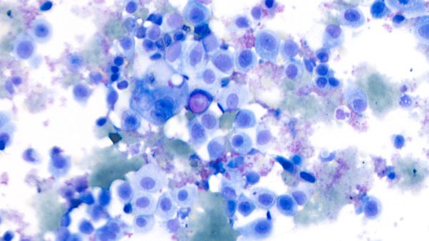
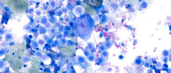
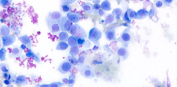
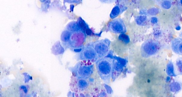
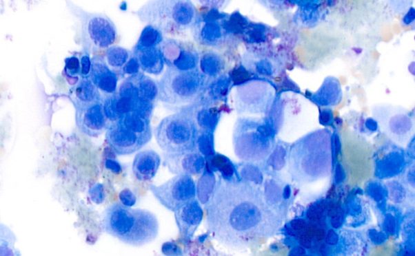
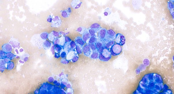
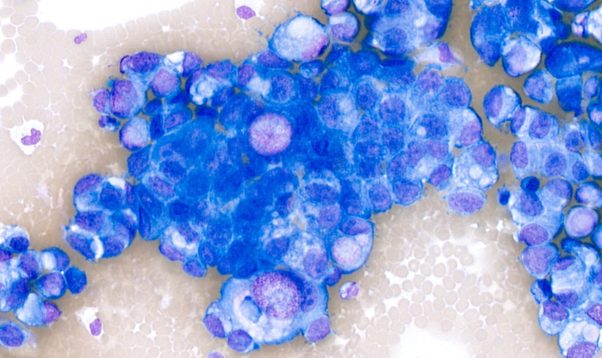
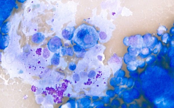
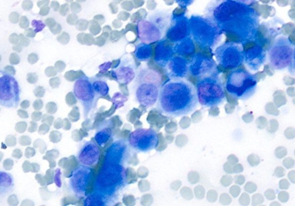
The predominant population consisted of round to polygonal epithelial cells with often distinct cell borders. The cells had round to oval, large nuclei with finely stippled chromatin, moderate to abundant basophilic cytoplasm, occasional prominent nucleoli and rare eosinophilic, rounded, intracytoplasmic inclusions (Melamed-Wolinska bodies). Anisocytosis and anisokaryosis were moderate to marked; rare, giant multinucleated cells were also present. Additionally, low numbers of segmented neutrophils and macrophages were noted.
These findings combined with the presence of Melamed-Wolinska bodies were highly suggestive of a urothelial carcinoma. The overrepresented breeds are Shetland sheepdogs, West Highland white terriers, fox terriers, and beagles. Studies suggest they are enlarged lysosomes occurring in degenerating urothelial cells. The eosinophilic, round, intracytoplasmic inclusions can be found within a benign or malignant urothelial cells. If they are found outside of the urinary tract, a metastatic urothelial carcinoma may be suspected. Histology is always considered a gold standard to confirm these findings.
Case 1
Case 2
Share this article






Follow us
Get Pathology News Delivered to Your Inbox