Author: Arturo Loaiza-Bonilla, MD, MSEd
Pathology services are critical — but the workforce is stretched thin. In the United States, roughly 6.4 pathologists per 100,000 people serve a population facing rising cancer incidence and growing diagnostic complexity. About 26.5% of US pathologists are aged 65 or older, leaving a mounting replacement gap.
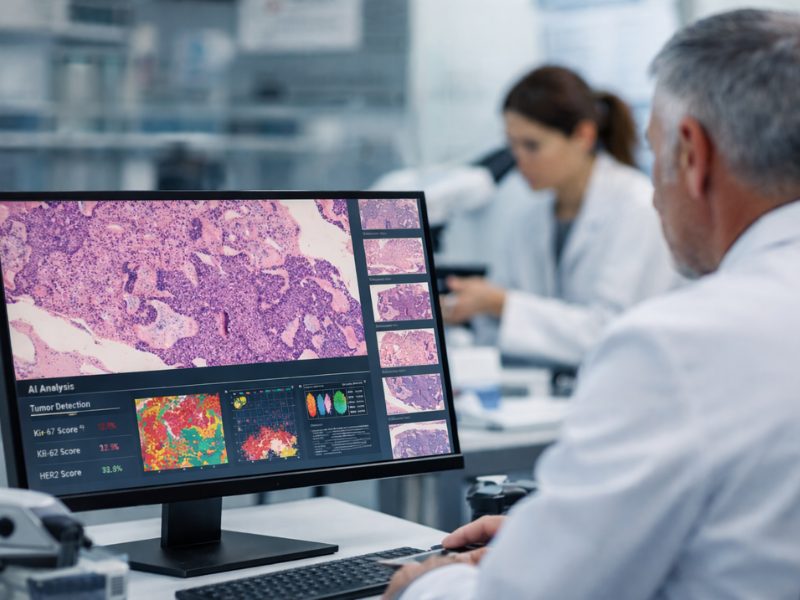
Globally, the imbalance is even more startling: In parts of sub-Saharan Africa there may be one pathologist per 1.5 million people and, in some low-resource countries, there may be no public-sector pathologists at all. In this context, digital pathology and artificial intelligence (AI) become more than conveniences; they become force multipliers by enabling remote review, sharing among experts across geographies, and automation of repetitive tasks — all of which ultimately help pathologists focus where their cognitive impact is highest.
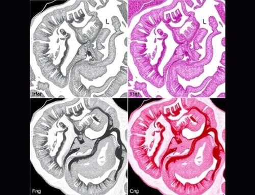
If radiology was AI’s first proving ground, pathology is its next. For decades, the microscope slide has been the bedrock of cancer diagnosis. Today, the slide isn’t just a piece of glass, it’s a computable dataset, and the lab bench is becoming a scalable AI platform. For oncology teams, that matters because beyond being a force multiplier, it is also a utility multiplier: The same tissue you’ve already collected can be reused to unlock prognostic and predictive signals that complement genomics.
Here’s what changed — and why this new data layer deserves a place in your clinic’s playbook.
Whole-Slide Imaging: The Essential Data Layer
A decade ago, digitizing slides was a convenience for remote consults. Now, in 2025, whole-slide imaging has become the foundational substrate that enables modern pathology AI.
In 2017, the FDA cleared the first whole-slide imaging system for primary diagnosis (Philips IntelliSite), removing a long-standing barrier to adoption.
Momentum accelerated in the past year or two, when Roche expanded its Digital Pathology Dx clearances to include both the VENTANA DP 200 and high-throughput DP 600 scanners, making routine primary diagnosis feasible in US labs and opened the door to AI at scale.
Alongside this, the validation playbook (ie, what “good” looks like) has taken shape. The College of American Pathologists published an updated guideline that many labs now follow as a practical roadmap to validate whole-slide imaging for diagnostic use, which includes case mix, intraobserver concordance, and re-review protocols. The numbers highlighted in this roadmap are memorable: at least 60 cases per application and a target of ~95% diagnostic concordance between whole-slide imaging and glass (with a weighted mean of ~95.2%). If your pathology partner cites this guideline, you’re on a solid runway for AI integration.
For oncologists, the significance is straightforward: Once slides are digitized reliably and at scale, pathology stops being a single, static point result and instead becomes a reusable, AI-computable signal. The same biopsy can inform triage, prognosis, and even treatment selection— without additional tissue handling.
Foundation Models Are the New Engines
Pathology has entered its foundation-model era, echoing the shift that transformed natural language AI and radiology. Rather than training narrow algorithms for a single task, companies and labs now pretrain very large encoders on diverse whole-slide imaging, allowing downstream tools to plug into these backbones with less labeled data and to generalize better across stains, scanners, and sites.
A concrete example of this isPLUTO-4 from PathAI. The PLUTO-4 family (including the compact PLUTO-4S and frontier-scale PLUTO-4G) was pretrained self-supervised on 551,164 whole-slide images from 137,144 patients across over 50 institutions, 60 diseases, and 100 stains. Evaluations show state-of-the-art transfer across patch-level, segmentation, and slide-level tasks, with double-digit improvement on a demanding dermatopathology benchmark — proof that pathology-specific pretraining is paying off.
From morphology to molecules, these encoders aren’t limited to cancer vs no cancer. Early results show they can predict spatial gene-expression signals from hematoxylin and eosin staining and capture biologically meaningful structure in their embeddings, which is key to building morpho-molecular biomarkers that complement genomics, computed directly from diagnostic slides.
The clinical takeaway is simple: Better backbones lead to more robust downstream tools. This is shown through improved calibration, fewer brittle failure modes across scanners and stains, and faster iteration from research to regulated tests.
Buildout Goes Beyond Any One Company
Two forces are accelerating the buildout of this new platform layer: massive data consolidation and clearer rules for remote digital practice.
In August 2025, Tempus acquired Paige, with the explicit goal of merging Paige’s roughly 7 million digitized slides with Tempus’ multiomic datasets to “build the largest foundation model in oncology.” Expect faster cycles from discovery to regulated tools as these corpora grow and as its models are retrained on ever-broader, better-labeled data.
Meanwhile, FDA-cleared whole-slide imaging stacks and image management systems are normalizing multi-AI workflows inside routine pathology, making it far simpler for your institution to pilot one AI module now and add another later without re-plumbing everything.
Cloud and Compliance
Cloud and compliance have entered a new phase, with regulators drawing bright lines that enable cloud-native, distributed pathology while closing public health emergency era loopholes. A Centers for Medicare & Medicaid Services revised memo confirms labs may continue remote review of digital data, results, and images (eg, whole-slide imaging viewed securely from home or another site) under the primary laboratory’s Clinical Laboratory Improvement Amendments (CLIA) certificate, if specified conditions are met.
However, the memo ends the public health emergency era flexibility that temporarily allowed for remote review of cytology digital slides without a separate CLIA certificate. This will end on March 23, 2026, after which labs will need separate certification for remote cytology after the transition period. This change does not impact remote review of surgical pathology whole-slide imaging. But make sure your pathology partners are aligning their policies and documentation accordingly. And just to clarify: Physical glass slides cannot be read remotely under the primary certificate. This regulatory clarity is a practical enabler for cloud-native workflows and distributed subspecialty input.
The bottom line is that validated whole-slide imaging+ stronger backbones + cloud compliance = AI that can scale across sites and service lines and allow subspecialists to weigh in wherever they are, all without shuttling glass or duplicating tissue.
What To Ask Your Pathology Partner This Month
With whole-slide imaging validated, foundation models maturing, and cloud rules clarified, the stage is set for clinically actionable AI — not just detection but also risk and treatment-benefit from the diagnostic biopsy itself.
Arturo Loaiza-Bonilla, MD, MSEd, is the co-founder and chief medical AI officer at Massive Bio, a company connecting patients to clinical trials using artificial intelligence. His research and professional interests focus on precision medicine, clinical trial design, digital health, entrepreneurship, and patient advocacy. Dr Loaiza-Bonilla serves as Systemwide Chief of Hematology and Oncology at St. Luke’s University Health Network, where he maintains a connection to patient care by attending to patients 2 days a week.
Credits
Medscape Oncology © 2026 WebMD, LLC
Any views expressed above are the author’s own and do not necessarily reflect the views of WebMD/Medscape or its affiliates.
No audio available for this article yet.
No quiz available for this article yet.