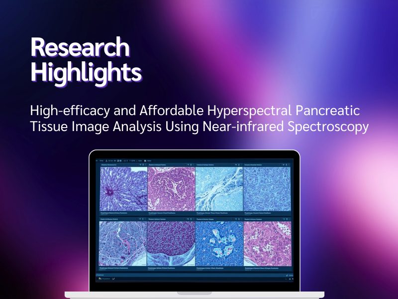
The following abstract is drawn from a recently published paper in Journal of Pathology Informatics. We invite you to read the full paper and join the conversation, become a member of the Pathology News community to share your thoughts, ask questions, and engage with others around this work.
Authors: Zheng Tanga, Abhinav Mishrae, Benjamin Moraa, Bilal Al-Sarirehb, Olivia Irvinee, Brandon Mauria, Victoria Higginbothamc, P.M. Anupama Bandaranayakee, S.H. Chandrashekharad, Venkat Kanamarlapudic, Debdulal Roye
Abstract
Pancreatic cancer remains one of the most lethal malignancies with less than 10% five-year survival rates, primarily due to late-stage diagnosis and limited early detection capabilities. Current diagnostic methods are expensive, time-intensive, and often inadequate for widespread screening applications. This study presents a novel, cost-effective approach using near-infrared (NIR) hyperspectral imaging combined with advanced machine learning for automated pancreatic tissue classification. We have developed a comprehensive pipeline incorporating autoencoder-based spatial feature extraction, multi-method consensus outlier detection, and systematically optimized neural network classifiers to distinguish between cancerous and non-cancerous pancreatic tissue samples. Our methodology was evaluated on 78 tissue microarray samples, with rigorous quality control yielding a final dataset of 69 high-quality specimens. The optimized classification model achieved 84% balanced accuracy using leave-one-out cross-validation, representing a 10% point improvement over conventional FICA+SVM approaches (74.0%) and approaching the performance of expensive conventional histopathological methods. Key technical innovations include consensus-based outlier detection, systematic hyperparameter optimization revealing optimal single-layer architectures with ELU activation, and interpretable attention mechanisms for diagnostic decision support. The demonstrated cost-effectiveness of NIR instrumentation combined with robust classification performance positions this approach as a promising pathway toward accessible, real-time pancreatic cancer screening tools that could significantly impact early detection rates and patient outcomes in diverse clinical settings.
Read the full article: High-efficacy and affordable hyperspectral pancreatic tissue image analysis using near-infrared spectroscopy – ScienceDirect
- aDepartment of Computer Science and Mathematics, Swansea University, Swansea SA2 8PP, UK
- bMorriston Hospital, Heol Maes Eglwys, Morriston SA6 6NL, UK
- cSwansea Medical School, Swansea University, Swansea SA2 8PP, UK
- dAll India Institute of Medical Sciences, Ansari Nagar, New Delhi 110029, India
- eDepartment of Chemistry, Swansea University, Swansea SA2 8PP, UK
No summary available for this article yet
No audio available for this article yet.
No quiz available for this article yet.