by Christos Evangelou, MSc, PhD – Medical Writer and Editor
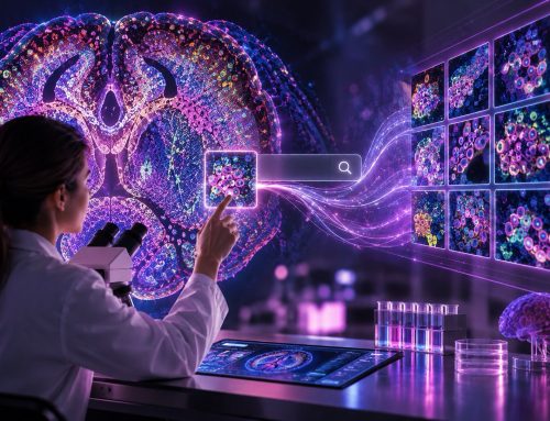
When CyTOF technology was first developed for analyzing cells in suspension, critics consistently pointed out the loss of tissue context as a key limitation. That criticism became the driving force behind one of the most transformative technologies in spatial biology. CODEX, developed in Nolan's Stanford laboratory, now allows researchers to analyze more than 50 protein markers simultaneously while preserving the spatial relationships between cells in cancer tissues. In an interview with Pathology News, Dr. Nolan discussed how spatial proteomics is helping researchers and pathologists map cellular neighborhoods and predict treatment outcomes. He also shared his insights into how artificial intelligence (AI) can further advance spatial proteomics analyses.
Origins and Evolution of CODEX
The journey from CyTOF to CODEX was not just about technological advancement — it was about enhancing our understanding of spatial cellular contexts within tumors.
"The vision was to take what we had done with CyTOF and address the criticism that we didn't know the actual context of cells in tissue," Nolan explained. "This was especially important for understanding the cancer-immune interface."
The transition from analyzing cells in solution to preserving their spatial relationships has proven to be more powerful than initially anticipated.
"Once you bump up to 50 or 70 markers, you get a chance to assay every single cell in the tissue," Nolan noted. "You may not know everything about them, but you know a lot more about what they are."
Mapping Cancer Cell Neighborhoods
One of the most important strengths of CODEX is that it allows researchers to study the complex spatial organization within tumors. Nolan's team discovered that it is not just which cell types are present, but how they are organized that determines patient outcomes. As he explained, in their colorectal cancer study published in Cell, they found that it was the size of the neighborhood, not the neighborhoods themselves, that predicted outcomes.
Their study showed that larger, more contiguous immune cell neighborhoods correlate with better patient outcomes, while fragmented neighborhoods — "like a broken mirror," as Nolan described — indicate worse prognosis. This suggests that tumors actively work to disrupt immune organization.
"The goal of the tumor is to disrupt organization, and the more the tumor disrupts organization, the worse the outcome," Nolan said.
Nolan noted that spatial interactions between cells are particularly complex at the boundaries between different cellular neighborhoods.
"Although we draw sharp boundaries in our papers, they're actually gray zones that merge into each other," he described. "When you look at cells in these merged zones, about 10 cells wide, the markers are drastically changed relative to cells on either side."
These boundary zones represent cells in transition, moving from one cytokine environment to another.
"You're looking at perturbations that you would love to have been able to create in tissue culture, but now it has all the cells and extracellular matrix present," Nolan explained.
Using CODEX to Study the Extracellular Matrix
In recent studies, researchers have combined matrix-assisted laser desorption/ionization mass spectrometry imaging with CODEX to identify cell-specific extracellular matrix structures.
"The extracellular matrix is not only the scaffold on which cells move, but it can also be a barrier that prevents the immune system from attacking the tumor," he explained. "We didn't realize there were this many types of extracellular matrix — we used to think of extracellular matrix as one thing, just like I used to think of macrophages as one thing."
Clinical Translation of Spatial Proteomics
The progression of CODEX from a research tool to a clinical application is already underway. Nolan emphasized that clinical implementation does not require the full complexity of research applications.
"You can't do 100 antibodies — it would break most laboratories' budgets. But through statistics, you figure out the minimal marker set that will be useful," he explained.
Current clinical applications are focusing on panels of around 10 markers that can provide actionable information within the timeframe required for patient care.
The company Akoya Biosciences, which commercializes CODEX technology, is already working with pharmaceutical companies to use spatial protein markers for patient stratification in clinical trials.
"It's not the 'boil the ocean' approach," Nolan noted, "but focused panels that can provide meaningful results."
Challenges of Implementation
Challenges limiting the widespread adoption of CODEX in research and clinical laboratories include cost considerations, the need for specialized equipment, and, most importantly, the computational expertise required for data analysis and interpretation. However, Nolan is optimistic about solutions to computational challenges and data analysis.
"There are now pipelines available that take the data all the way from original output to analysis," he explains.
He gave Cellformatica as an example of an AI platform for analysis of high-throughput data, noting that
"It's all done on the cloud with no special software or local servers needed."
For researchers starting with CODEX, Nolan advocates for a pragmatic approach.
"I always push the 95% rule in the lab. If you've got 95% of what you wanted, go with it. Don't spend another six months chasing that last 5%."
He recommended starting with smaller antibody panels to "burn the pipeline" and learning the technical aspects before attempting larger, more complex studies.
The Application of AI in Spatial Biology
Nolan sees AI as the key to enhancing the impact of spatial proteomics technologies.
"Large language models are going to tell us about marker-level differences we didn't appreciate were in the data," he predicts. "They'll point to marker sets we could not have thought of previously."
His vision of using AI extends beyond data analysis.
"AI is going to tell us more about what markers we should be using as diagnostics, as opposed to just the ones we're figuring out at a high level. It will interpolate the best markers for us."
He uses the metaphor of a spider's web:
"The diagnostic we're doing is like a web of changes. The farther the fly is from the spider, the harder it is to find it. But if AI knows everything about the web, it can figure out markers closer to the originating cause."
Integration Across Data Types
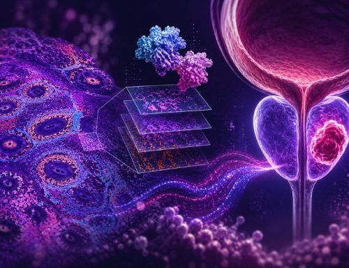
The MaxFuse algorithm, developed in Nolan's laboratory, is another step forward in spatial biology. By integrating protein and RNA data, researchers can now see previously hidden diversity within seemingly uniform cell populations.
"By protein alone, a germinal center would all be blue — just B cells. But by integrating RNA data, we now see all these differences that nobody appreciated previously," Nolan explained.
This multimodal integration works because different data types — RNA, protein, and others — are essentially different "languages that speak about the cell." The relationships between these languages allow for translation between data types, similar to how language translation algorithms work.
For pathologists and researchers who are hesitant about studying spatial proteomics, Nolan encouragingly said, "Jump in — the water is not that deep." He particularly encourages students and early-career researchers not to be deterred by established opinions.
As Nolan concluded, understudying cancer pathology lies not just in identifying cell types but also in mapping the spatial relationships that determine how tumors grow, evade immune responses, and respond to treatment. CODEX and other spatial technologies are providing the tools to read these spatial stories, one neighborhood at a time.
No audio available for this article yet.
No quiz available for this article yet.