by Christos Evangelou, MSc, PhD – Medical Writer and Editor
New research using silicon nanoneedles has provided insights into the molecular evolution of living brain tissue. According to the authors, this method creates “molecular replicas” that capture the lipid landscape of brain tumors without damaging the tissue, offering clinicians a potential tool for understanding how gliomas evolve and respond to treatment in real time.
The study was published in Nature Nanotechnology.
Study Rationale
Traditional spatial biology technologies rely on fixed, non-living tissue samples. This makes it challenging to track how molecular profiles change over time within the same specimen.
"We saw a clear clinical need to understand how tissue microenvironments evolve over time through dynamic molecular regulation," explained Dr. Ciro Chiappini, the corresponding author of the study. "This information is crucial for identifying emerging resistance pathways or mechanisms of disease progression, but it's extremely difficult to obtain using traditional fixed-section analyses."
The research team, led by Dr. Chiappini and colleagues from institutions across the UK and Israel, developed a method that uses porous silicon nanoneedles to repeatedly sample biomolecules from live brain tissue. The researchers use this method to conduct lipid analysis and assess how metabolic markers are altered in gliomas during treatment.
Nanoneedle Technology
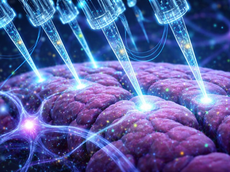
The system contains arrays of porous silicon nanoneedles, each measuring 4 micrometers in height with 50-nanometer tips, which are briefly applied to tissue surfaces for approximately 10 seconds. During this contact, biomolecules transfer from the tissue to the nanoneedle substrate, creating what the researchers term a "molecular replica."
"The process is simple and minimally disruptive," Dr. Chiappini emphasized. "We assessed tissue viability after sampling and found no difference compared to unsampled controls, even after 5 days, confirming that the procedure is non-destructive."
The nanoneedles have a surface area of 77.2 m²/g and 50% porosity, and their porous structure is essential for effective biomolecule collection. The researchers found that porous nanoneedles significantly outperformed solid alternatives, capturing more molecular features with greater reproducibility.
Validation of Molecular Replicas
The team conducted validation studies to compare the lipidomic profiles of the replicas with those of their corresponding tissue sections and found that the nanoneedle replicas accurately represent the molecular composition of the tissue.
The correlation factor exceeded 0.95 across a wide range of lipid abundances, and repeated sampling from the same tissue yielded correlations above 0.9.
"These results confirm that our approach provides a highly representative snapshot of the molecular state of the tissue," Dr. Chiappini stated.
The molecular replicas captured morphological features of the brain and were able to distinguish between gray matter, white matter, and tumor regions based on their distinct lipid signatures. Hierarchical clustering analysis showed that replicas could identify the same tissue regions as traditional sections, with white matter characterized by high levels of sulfated hexosylceramide and gray matter distinguished by elevated phosphatidylserine levels.
Clinical Validation and Disease Classification
The researchers analyzed 23 human glioma biopsies using both traditional tissue sections and nanoneedle replicas to assess the clinical relevance of the method. Machine learning analysis demonstrated that replicas could classify samples based on disease state with comparable accuracy to conventional approaches, achieving an area under the curve value of 0.75 ± 0.02 for replicas versus 0.71 ± 0.05 for tissue sections.
The research team also identified lipid biomarkers associated with tumor grade. Phosphatidylethanolamine and phosphatidylserine species were associated with low-grade tumors, whereas phosphatidylinositol and other phosphatidylserine variants were linked to high-grade disease. Additionally, the spatial distribution of these markers aligned with expected tumor grades and captured intratumoral heterogeneity.
Spatiotemporal Analysis Reveals Treatment Dynamics
The team conducted longitudinal studies of living glioma tissue slices treated with temozolomide, sampling the same tissue at multiple timepoints to track how lipid profiles evolved in response to treatment.
This analysis revealed both time-dependent and treatment-specific changes in lipid composition. At the molecular level, temozolomide treatment led to significant decreases in specific phosphatidylserine and phosphatidylinositol species, including the complete disappearance of phosphatidylserine 29:0 in treated tumors.
"One particularly interesting finding was the degree of sample-specific response," Dr. Chiappini noted. "When analyzing longitudinal samples from the same tissue, we could detect variations that would have been lost had we relied on separate samples for each time point. This highlights the value of a repeated, minimally invasive sampling approach."
Potential Clinical Translation
Dr. Chiappini envisions that their method can have several clinical applications, including screening precancerous lesions and providing intraoperative guidance during glioma surgery through real-time molecular analysis.
"One potential application is in screening accessible precancerous lesions, such as those in the oral cavity that may develop into head and neck cancers," Dr. Chiappini explained. "Our approach could allow molecular monitoring of such lesions without the need for a biopsy, improving early detection."
The technology can be incorporated into existing medical devices, such as endoscopes, enabling access to diverse anatomical sites for molecular sampling, he added.
Limitations and Future Directions
Limitations of the method include its focus on lipidomics, although expansion to other molecular classes is underway. Additionally, the spatial resolution may need to be enhanced for single-cell applications.
"A key focus is expanding beyond lipidomics to integrate spatial transcriptomics and proteomics, which are currently under development in our lab," Dr. Chiappini noted.
The team is also working to standardize data interpretation protocols and ensure reproducibility across different clinical settings.
Dr. Chiappini emphasized that with future optimization, the method's potential could extend beyond glioma research.
"The ability to sample live tissue non-destructively has broad potential, and we are actively exploring its use in other tissue types and disease models," he said.
References
- Gu C, Martella DA, Rose LA, et al. Nanoneedles enable spatiotemporallipidomicsof living tissues. Nat Nanotechnol. Published online June 16, 2025. doi:10.1038/s41565-025-01955-8
No audio available for this article yet.
No quiz available for this article yet.