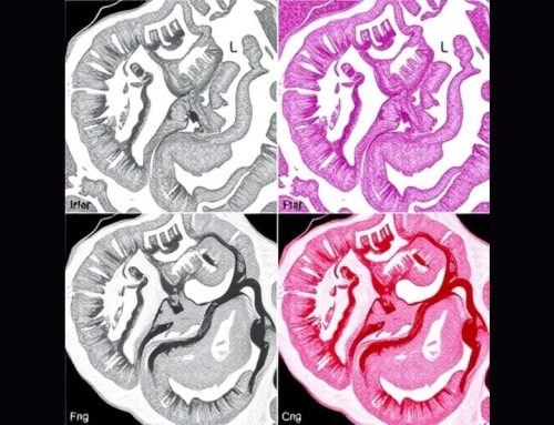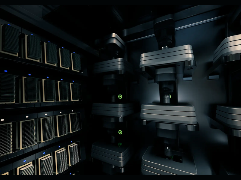
The Tissue Image Analytics (TIA) Centre at the University of Warwick has announced the release of TIAToolbox v2.0.0, a major update to its open‑source platform for digital pathology image analysis. Designed to meet the growing needs of pathologists, researchers, and clinical teams, TIAToolbox provides end‑to‑end tools for working with whole‑slide images (WSIs), running AI models, and visualising results in ways that integrate naturally into diagnostic and research workflows.
What is TIAToolbox?
TIAToolbox is an open‑source software library designed to help researchers analyse whole‑slide images (WSIs) using modern artificial intelligence (AI) techniques. It provides ready‑to‑use tools for tasks such as cell and nucleus detection, tissue segmentation, patch extraction and feature analysis, while also supporting custom workflows for more advanced research. Importantly, the toolbox aims to make computational pathology approachable for users with varied levels of technical expertise from trainees exploring digital slides, to researchers building large‑scale studies.
Why This Release Matters for Pathologists
Digital pathology continues to expand rapidly as departments adopt whole‑slide scanners and AI‑assisted diagnostic workflows. Modern scanners routinely produce WSIs containing billions of pixels per slide, and analysing these images remains a major bottleneck for laboratories and research teams.
TIAToolbox v2.0.0 has been engineered to make this analysis significantly faster, more reliable, and easier to incorporate into real‑world clinical and research workflows.
What’s New in This Release?
A Fully Redesigned Engine for Modern AI Workflows
Version 2.0.0 introduces a complete redesign of the deep‑learning engine at the core of TIAToolbox. This new engine has been built from the ground up to meet the needs of contemporary pathology AI:
- Speed: In benchmarking studies, the new engine delivered dramatic performance gains. On the widely referenced CMU‑1.ndpi whole‑slide image (50k × 31k pixels), processing was approximately 25× faster than in previous versions, with even greater gains observed on larger slides.
- Scalability: Designed for gigapixel‑scale whole‑slide images, the system handles high‑resolution data more efficiently, making large retrospective cohorts and clinical research studies more feasible.
- Ease of use: Despite the technical advances, the user interface remains consistent and streamlined. Users can switch between output formats or models without needing to rewrite workflows.
To hear more about the work behind TIAToolbox and the TIA Centre, listen to our recent podcast conversation with Dr Shan Raza, where we discuss the growth and mission of the centre, the development of open-source tools for digital pathology, and how multimodal data, international collaboration, and grand challenges are shaping the future of AI in medicine.
Conversation with Dr Shan Raza, TIA Centre at Warwick University – Pathology News
No audio available for this article yet.
No quiz available for this article yet.