One Integrated Breast AI Solution
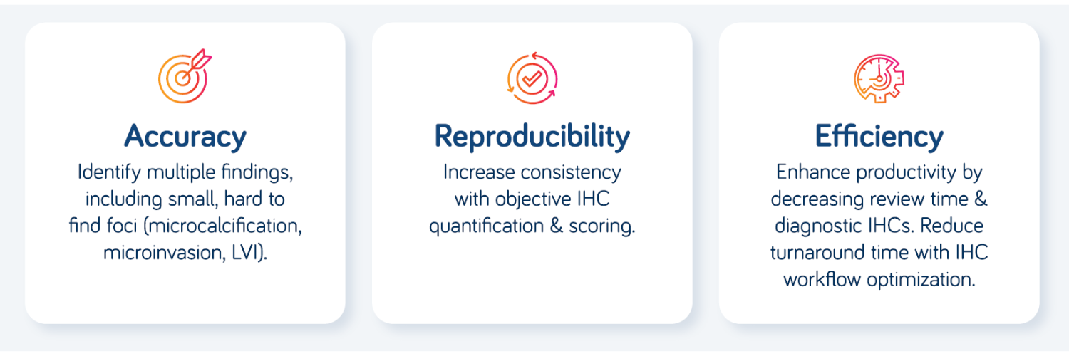
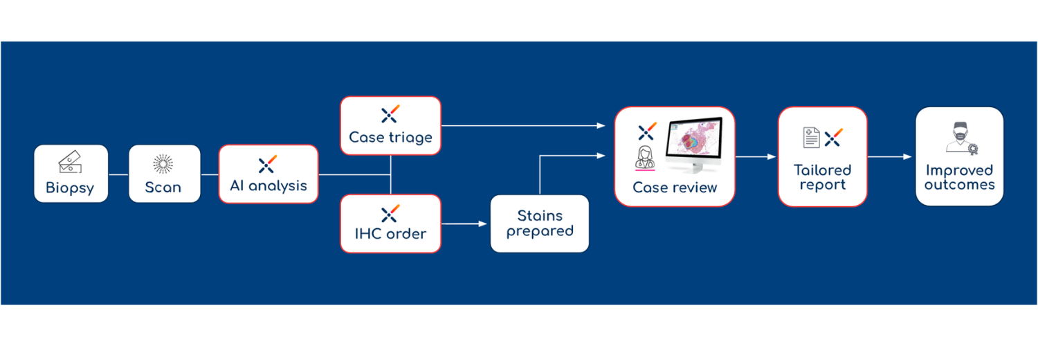
Replace fragmented tools with Ibex Breast, a single platform that simplifies your workflow and reduces review times up to 43%.6
Ibex Breast AI Models
Ibex Breast H&E
Precision AI for Breast Pathology
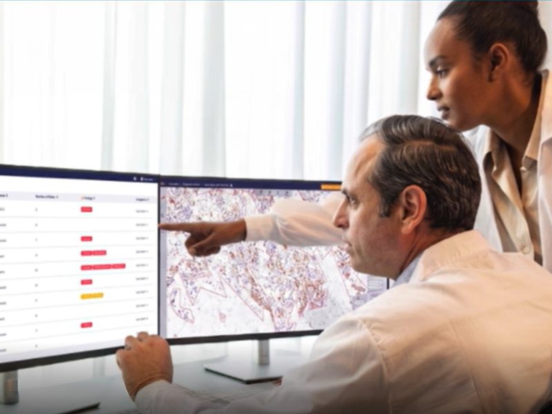
Automate the Search, Focus on the Patient’s Diagnosis:
- Detects >50 malignant, pre-malignant & benign tissue morphologies (microinvasion, microcalcification, LVI, IDC vs ILC, DCIS grading, lobular neoplasia, TILs and more1-11)
- Eliminates tedious manual reviews
- Optimizes IHC workflows by pre-screening for invasive cancer
- The AI is exceptionally robust, with high performance across multiple labs, scanners and patient demographics.1-11
Ibex Breast HER2
Accurate, Reproducible HER2 Scoring for Targeted Therapies: Identify HER2-low and -ultralow cases with confidence.
Ibex Breast HER2 uses AI to automate 2023 ASCO/CAP scoring guidelines, removing subjectivity and ensuring reproducible precision for every patient, with:
- “Zero-click” invasive tumor detection (automatically excluding ductal carcinoma in situ)
- Automated tumor cell identification and cell staining pattern classification
- Interactive invasive contours and score recalculation
- Visualization of AI findings and quantification of cell staining pattern percentages for better understanding of score result
The AI is exceptionally robust, with high performance across multiple labs, scanners and patient demographics.12-17
Ibex Breast ER/PR
Precision ER/PR Scoring for Every Patient.
Ibex Breast ER/PR delivers automated ER/PR quantification, delineating expression into positive and negative based on ASCO/CAP 2020 guidelines, ensuring every patient is accurately identified for optimal endocrine therapy, with:
- “Zero-click” invasive tumor detection (automatically excluding DCIS)
- Automated tumor cell identification and cell staining pattern classification
- Interactive invasive contours and score recalculation
- Visualization of AI findings and quantification of cell staining pattern percentages for better understanding of score result
Ibex Breast Ki67
Objective, Automated Ki67 Quantification: Replace manual counting with AI-powered precision.
Aligned with International Ki67 Working Group and St. Gallen guidelines, Ibex Breast Ki67 provides “zero-click” quantification for accurate, reproducible patient stratification, with:
- “Zero-click” invasive tumor detection (automatically excluding ductal carcinoma in situ)
- Automated tumor cell identification and cell staining pattern classification
- Interactive invasive contours and score recalculation
- Visualization of AI findings and quantification of cell staining pattern percentages for better understanding of score results.
References:
- Lami et al. Pathology (2024), 56; 5:633-642
- Tahir et al. Clin Breast Cancer. 2025
- Sandbank et al. NPJ Breast Cancer 2022
- Vincent-Salomon et al. Modern Pathology 2022, 35 (Suppl 2): 203-204
- Vincent-Salomon et al. Virchows Arch. 2022. 481 (Suppl 1), S48
- Vecsler et al. Virchows Arch. 2025 Sep;487(Suppl 1):PS-01-038
- Broeckx et al. Virchows Arch (2023) 483 (Suppl 1): S36
- Aslam et al. Virchows Arch (2023) 483 (Suppl 1): S58
- Karakas et al. Laboratory Investigation (2024), 104;3: S184-S185
- Sandbank et al. Modern Pathology 2022, 35 (Suppl 2): 1094
- Canas-Marques et al. Virchows Arch. 2025 Sep;487(Suppl 1):OFP-05-007.
- Cyrta et al. ESMO Open (2024), 9; 54: 103071
- Krishnamurthy et al. JCO 42, e15150-e15150(2024)
- Krishnamurthy et al. JCO Precis Oncol 8, e2400353(2024).
- Krishnamurthy et al. Clin Cancer Res (2025), 31 (12_S): P1-07-03.
- Grinwald et al. Laboratory Investigation (2025), Volume 105, Issue 3, 102380
- Vecsler et al. Presented at SABCS 2025
- Internal clinical validation study data; pending publication
- Cyrta et al. Presented at ECDP 2025