BxAlign®
Key Benefits & Facts:
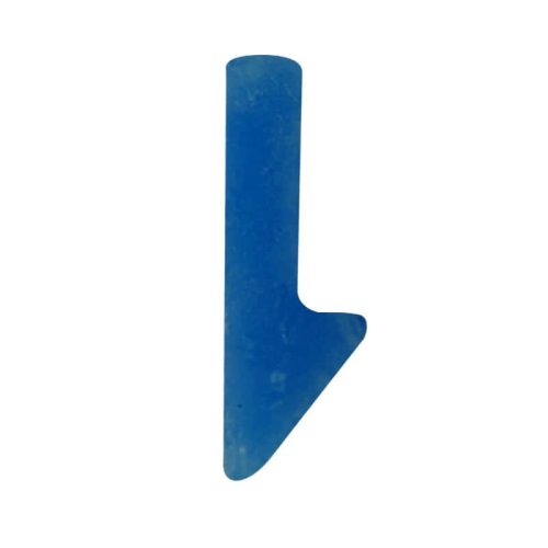
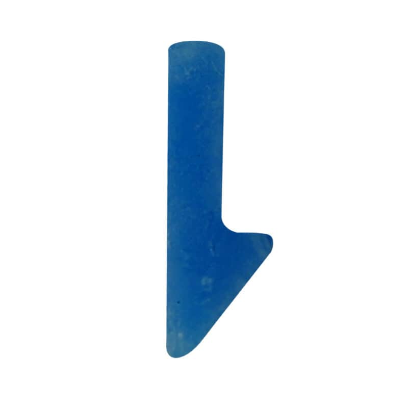
- Reduces scanning failures and rescans by providing a consistent plane of focus via the arrow.
- Serves as a fiducial marker for Lumea’s digital pathology software and AI, ensuring consistent positioning across all sections.
- Allows pathologists to view multiple levels and stains of the same specimen at once, enabling easier comparison.
- Compatible with standard lab processes, including staining and digitizing.
- One BxAlign block provides up to 100 sections when cut at 3 to 4 microns thick on the micrometer
To use, lab personnel section the BxAlign block at the same thickness as the tissue or cell structure that will go with it, deparaffinize and clear it on a slide, place the desired sample next to it on the slide, and then they can resume standard processing for staining and digitizing. Once uploaded into Lumea’s Viewer+™ or BxLink™ it can be used with Lumea’s MultiView for optimal digital slide comparisons and review.
Contact Lumea for more information on how BxAlign can enhance your lab’s efficiency and accuracy and for more specifics on how it works.
More Solutions from Lumea
Compare Solutions
Get Pathology News Delivered to Your Inbox
Learn More
Lumea News
Innovators at the Table: Hamamatsu’s Path from Photons to Pathology
Editor2026-07-24T15:03:01+00:00July 21, 2026|
Lumea Receives Global Recognition Award for Exceptional Achievement
Editor2026-06-30T08:25:40+00:00June 26, 2026|
One Dermatopathologist’s Case for Going Digital – Podcast
Editor2026-06-23T15:49:28+00:00June 18, 2026|
Your Digital Pathology Adoption: Separating Essential Gear from Digital Dead Weight
Editor2026-06-17T15:10:05+00:00June 15, 2026|
Lumea Named Best New Biopsy Technology Solution in 2026 MedTech Breakthrough Awards
Editor2026-05-19T10:40:33+00:00May 15, 2026|
Announcing the DDx Pathology Foundation Scholarship!
Editor2026-05-18T12:45:13+00:00May 13, 2026|
Digital Diagnostic Summit Launches Specialty-Specific Educational Series to Accelerate Innovation in Prostate Cancer Diagnostics
Editor2026-05-05T10:24:33+00:00April 24, 2026|


Reviews
There are no reviews yet.