Author: Sidney Ocanagil-Tunstall
In a pathologist’s office, a stack of glass cases carries information beyond the slides themselves. It shows what needs attention. A case set aside can serve as a reminder that an additional stain is pending. Removing that stack means finding another way to communicate what is ready, what is missing and what needs following up.
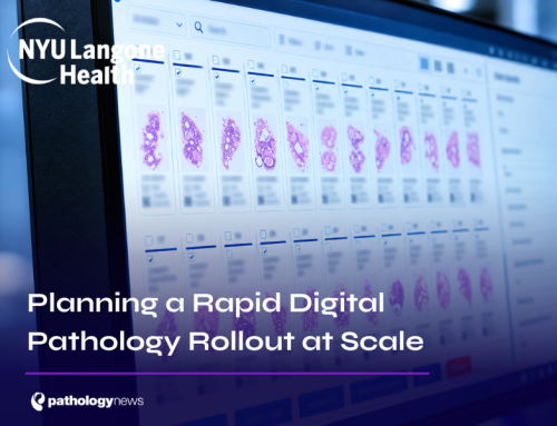
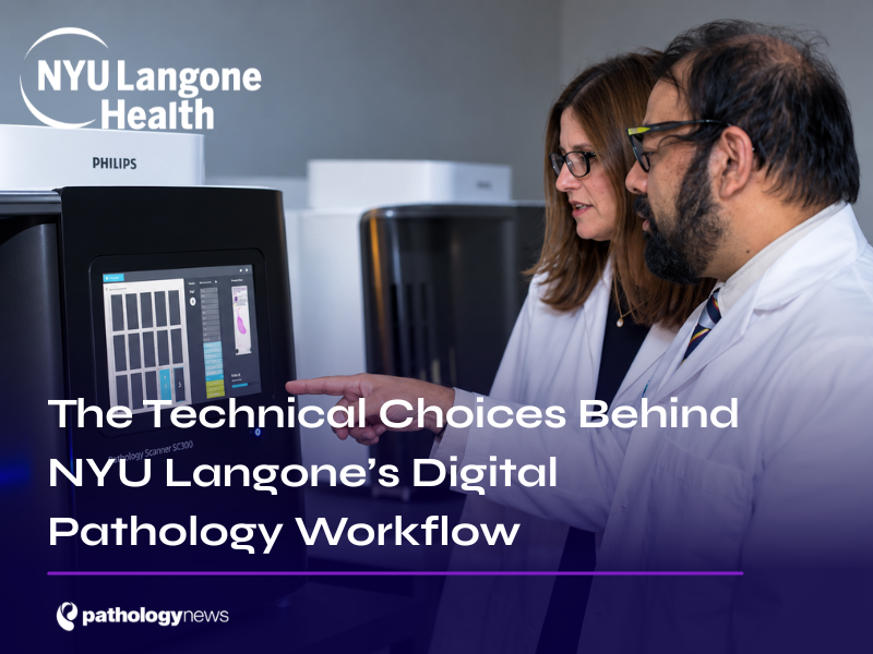
During our Key Labs visit to NYU Langone, Dr Syed T. Hoda, Director of Digital Pathology demonstrated how the team had addressed this through its existing Epic Beaker laboratory information system. Worklist indicators showed whether laboratory tasks were complete and images were available. Ordering a stain changed the case’s status; completing and scanning the additional work changed it back.
Behind that apparently simple interaction was a deliberate technical choice: customise the LIS, EPIC Beaker around NYU’s digital workflow and connect it to the imaging environment. Amin El-Rowmeim, Senior Manager, Clinical Ancillary Systems at NYU Langone Health, explains how that integration, and the infrastructure supporting it, took shape.
Building the workflow with the LIS in mind
NYU’s pathologists already signed out reports in Epic Beaker. The team retained that environment, although Amin says it could not find an Epic module for digital pathology at the time.
“We did keep the pathologist in Epic Beaker because they’re already signing reports in Epic Beaker,” he explains.
NYU developed a custom integration linking Beaker to its image management system, Philips IntelliSite Pathology Solution. Pathologists could open images directly from the case without a further login. Hoda describes removing that repeated interruption as an intentional part of the design.
The worklist addressed a more substantial problem. Hoda explains that pathologists had rarely used it before, because they could scan a physical slide’s identifier to bring up the patient. With glass distribution ending, the software needed to make case readiness visible.
In his demonstration, a monitor icon indicated that images were scanned and ready. A yellow indicator warned that not everything was available. Separate indicators showed whether laboratory tasks were complete and whether the pathologist had ordered additional work. Together, these could distinguish a completed laboratory task from a slide still awaiting scanning.
“This requires IT to build two-way communication,” Hoda explains. An additional order created a placeholder in Philips; as the corresponding scanned material became available, the status updated. He recalls that building this simplicity took months.
Customisation also provided visibility into adoption. A question recorded the method of slide review for each case, distinguishing digital, digital-plus-glass and glass-only review during the transition. Those responses supplied adoption metrics and informed the report disclaimer. The system could therefore support daily work while giving the implementation team evidence of how it was being used.
Keeping the imaging infrastructure manageable
Amin describes a further decision beneath that clinical interface. NYU was initially advised to use multiple IMS servers. After working through the recommendation with its vendor, it adopted a single enterprise IMS arrangement, avoiding duplicated interfaces and infrastructure. A secondary data centre provided a separate recovery environment.
Achieving the integration required NYU’s LIS and cloud specialists to work alongside suppliers. Hoda’s broader account of the rollout reinforces the importance of that coordination: project management brought parallel clinical, technical and administrative work together, while the implementation was adapted to NYU’s own staffing and case-handling practices.
Retaining Beaker gave the project an established reporting environment to build upon. The custom work then supplied information that pathologists would lose when their cases stopped arriving as physical slides.
Making images responsive at scale
“The goal is to make the digital pathology platform exactly like a microscope,” says Amin.
Pixelation and delays during panning and zooming were significant sources of dissatisfaction to avoid. NYU deployed dedicated high-speed network equipment, kept diagnostic workstations on wired connections and used high-end workstations with GPUs to render images locally.
Performance was tested under increased demand. Amin describes doubling the number of scanners during stress testing to assess viewing at peak load. Ongoing monitoring tracked network performance against established baselines and checked that workstations remained wired.
Remote reporting required further refinement. NYU’s remote connectivity used Cisco Meraki MX SD-WAN, and Amin credits the later introduction of a hardware VPN with a substantial speed improvement. At the time of the interview, he reported that remote sign-out had risen from approximately 6% to 31%, with 57 pathologists reading remotely.
Controlling the data entering the archive
At approximately 6,000 whole-slide images archived each day, decisions about image size had lasting consequences. NYU used compression and tissue detection algorithms to bring average image size to approximately 750 MB.
“We actually started by looking at the whole slide image by avoiding storing unnecessary data,” Amin explains.
Images then moved through storage tiers. NYU’s documented configuration used Dell EMC PowerMax SAN storage for initial ingestion, followed by Dell EMC Isilon/PowerScale NAS storage. Amin describes that first transfer taking place overnight. Signed-out case images subsequently moved into AWS cloud storage, using S3 Standard and then Glacier archive tiers.
The strategy matched rapid access for active work with slower retrieval for archived material. Amin describes immediate retrieval from warm storage and approximately five to 12 hours from cold storage—still favourable, in his account, to retrieving physical slides from NYU’s archive in New Jersey.
After 22 months, the archive contained more than two million slides, occupying 1.67 petabytes. Some 77% of the data was already in the cloud. Moving older images off premises limited the hardware burden NYU would otherwise have to maintain as the archive grew.
Testing the service and its fallback
NYU began at its highest-volume hospital, where strong IT support was available, to validate scanner throughput and the enterprise workflow. Preparation included construction work to maintain suitable temperature and humidity in scanner rooms. Resolving technical and workflow challenges there made expansion easier, Amin recalls.
The team subsequently refined failover between its primary and secondary data centres so scanners could switch to the recovery environment without individual reconfiguration.
Amin describes digital pathology as a Tier 1 system, with enterprise monitoring across its servers, network and other components. Alongside backups, NYU replicated data to a secondary site containing an IMS and storage. Its recovery arrangements were exercised for an extended period each year.
“We’re mandated by leadership that we have to work from the secondary data center every year for about two to three weeks,” he explains.
That requirement meant the recovery environment had to support actual operation. It gave resilience a practical test beyond the existence of a documented plan.
The interoperability compromise
NYU also accepted a limitation. Hoda explains that the organisation prioritised FDA-cleared solutions when selecting its technology. Amin describes the resulting proprietary image format candidly: “It does have a proprietary image format … so we are in that locked-in state.”
For researchers, including those developing AI applications, NYU used the vendor’s software development kit to convert images at scale into DICOM or TIFF. Amin is explicit that this pathway was for research.
The longer-term plan involved DICOM-capable scanners and upgrades. At the time of the interview, NYU expected to receive scanners for testing, including assessment of clinical image quality, ahead of regulatory approval. Clinical DICOM capability remained a development goal.
The clearest expression of NYU’s technical approach is still the worklist Hoda demonstrated. Its simplicity depended on communicating orders, laboratory progress and image availability between systems. Storage, network performance and recovery arrangements then supported the pathologist’s ability to act on that information.
For a pathology lead, this is a concrete example of the work involved in making a digital service usable: identify what staff need to know at each step, build those signals into their working environment, and test the infrastructure they will depend upon. NYU’s Beaker customisation made that connection between technical design and everyday diagnosis visible.